


 Foundational characteristics of cancer include proliferation, angiogenesis, migration, evasion of apoptosis, and cellular immortality. Find key markers for these cellular processes and antibodies to detect them.
 The SUMOplot™ Analysis Program predicts and scores sumoylation sites in your protein. SUMOylation is a post-translational modification involved in various cellular processes, such as nuclear-cytosolic transport, transcriptional regulation, apoptosis, protein stability, response to stress, and progression through the cell cycle.
 The Autophagy Receptor Motif Plotter predicts and scores autophagy receptor binding sites in your protein. Identifying proteins connected to this pathway is critical to understanding the role of autophagy in physiological as well as pathological processes such as development, differentiation, neurodegenerative diseases, stress, infection, and cancer.
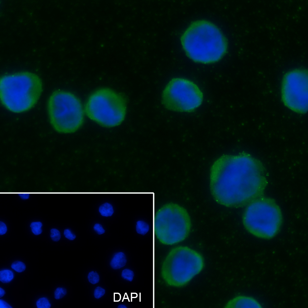
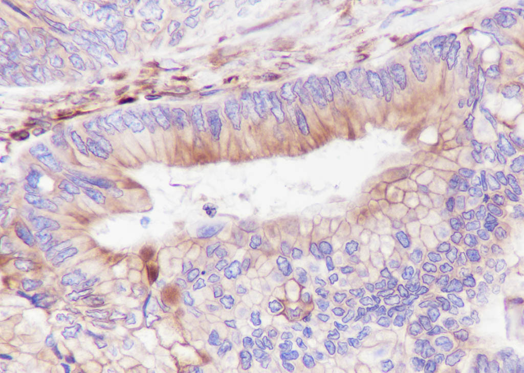
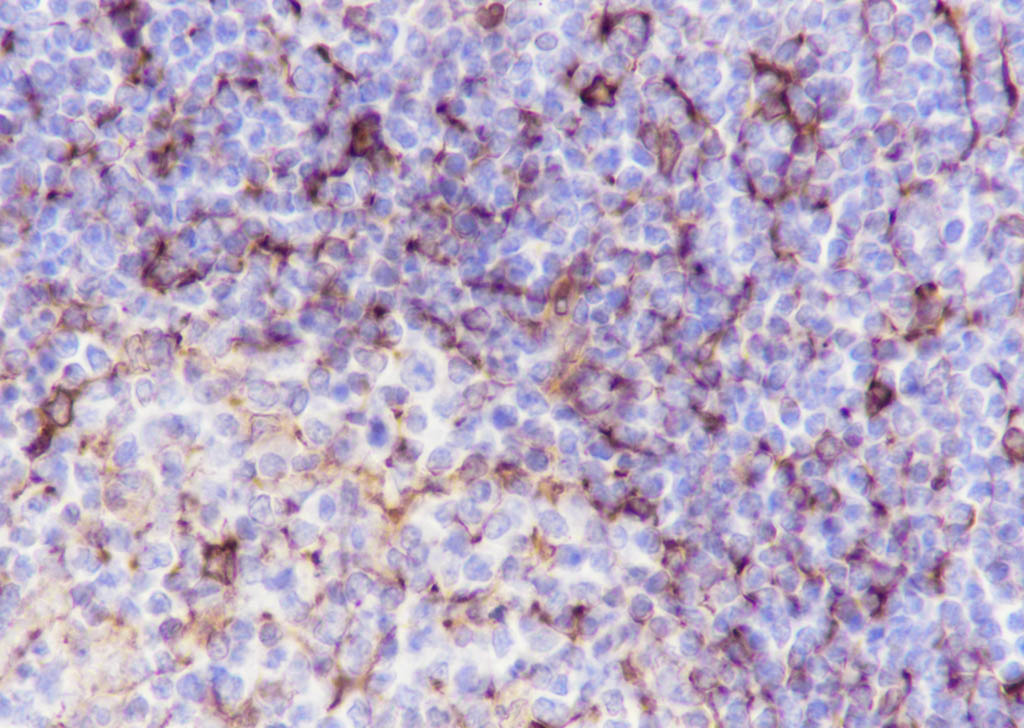
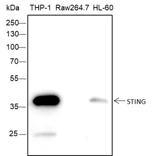
STING1 Recombinant Rabbit mAb
STING1 Recombinant Rabbit mAb


- SPECIFICATION
- CITATIONS
- PROTOCOLS
- BACKGROUND

Application
| WB, IHC-P, IHC-F, IF, ICC |
|---|---|
| Host | Rabbit |
| Clonality | Recombinant |
| Physical State | Liquid |
| Immunogen | A synthesized peptide derived from human STING |
| Epitope Specificity | 250-379/379 |
| Isotype | IgG |
| Purity | affinity purified by Protein A |
| Buffer | 0.01M TBS (pH7.4) with 1% BSA, 0.02% Proclin300 and 50% Glycerol. |
| SUBCELLULAR LOCATION | Endoplasmic reticulum membrane. Mitochondrion outer membrane. Cell membrane. Cytoplasm > perinuclear region. In response to double-stranded DNA stimulation, relocalizes to perinuclear region, where the kinase TBK1 is recruited. |
| SIMILARITY | Belongs to the TMEM173 family. |
| SUBUNIT | Associates with the MHC-II complex (By similarity). Homodimer; 'Lys-63'-linked ubiquitination at Lys-150 is required for homodimerization. Interacts with DDX58/RIG-I, MAVS and SSR2. Interacts with RNF5 and TRIM56. Interacts with TBK1; when homodimer, leading to subsequent production of IFN-beta. Interacts with IFIT1 and IFIT2. |
| Post-translational modifications | Phosphorylated on tyrosine residues upon MHC-II aggregation (By similarity). Phosphorylated on Ser-358 by TBK1, leading to activation and production of IFN-beta. Ubiquitinated. 'Lys-63'-linked ubiquitination mediated by TRIM56 at Lys-150 promotes homodimerization and recruitment of the antiviral kinase TBK1 and subsequent production of IFN-beta. 'Lys-48'-linked polyubiquitination at Lys-150 occurring after viral infection is mediated by RNF5 and leads to proteasomal degradation. |
| Important Note | This product as supplied is intended for research use only, not for use in human, therapeutic or diagnostic applications. |
| Background Descriptions | This gene encodes a five transmembrane protein that functions as a major regulator of the innate immune response to viral and bacterial infections. The encoded protein is a pattern recognition receptor that detects cytosolic nucleic acids and transmits signals that activate type I interferon responses. The encoded protein has also been shown to play a role in apoptotic signaling by associating with type II major histocompatibility complex. Mutations in this gene are the cause of infantile-onset STING-associated vasculopathy. Alternate splicing results in multiple transcript variants. [provided by RefSeq, Sep 2014] |
| Target/Specificity | Ubiquitously expressed. |
|---|---|
| Dilution | WB=1:500-1:1000,IHC-P=1:100-1:500,IHC-F=1:100-500,ICC/IF=1:20-50,IF=0 |
| Storage | Store at -20 ℃ for one year. Avoid repeated freeze/thaw cycles. When reconstituted in sterile pH 7.4 0.01M PBS or diluent of antibody the antibody is stable for at least two weeks at 2-4 ℃. |

Thousands of laboratories across the world have published research that depended on the performance of antibodies from Abcepta to advance their research. Check out links to articles that cite our products in major peer-reviewed journals, organized by research category.
info@abcepta.com, and receive a free "I Love Antibodies" mug.

Provided below are standard protocols that you may find useful for product applications.
Background
This product as supplied is intended for research use only, not for use in human, therapeutic or diagnostic applications.
If you have used an Abcepta product and would like to share how it has performed, please click on the "Submit Review" button and provide the requested information. Our staff will examine and post your review and contact you if needed.
If you have any additional inquiries please email technical services at tech@abcepta.com.
Ordering Information
Shipping Information
