


 Foundational characteristics of cancer include proliferation, angiogenesis, migration, evasion of apoptosis, and cellular immortality. Find key markers for these cellular processes and antibodies to detect them.
 The SUMOplot™ Analysis Program predicts and scores sumoylation sites in your protein. SUMOylation is a post-translational modification involved in various cellular processes, such as nuclear-cytosolic transport, transcriptional regulation, apoptosis, protein stability, response to stress, and progression through the cell cycle.
 The Autophagy Receptor Motif Plotter predicts and scores autophagy receptor binding sites in your protein. Identifying proteins connected to this pathway is critical to understanding the role of autophagy in physiological as well as pathological processes such as development, differentiation, neurodegenerative diseases, stress, infection, and cancer.
Hrasls3 Rabbit pAb
Hrasls3 Rabbit pAb
- SPECIFICATION
- CITATIONS
- PROTOCOLS
- BACKGROUND

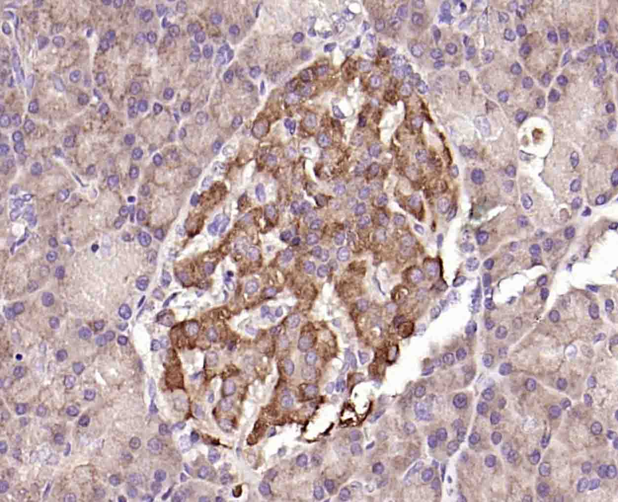
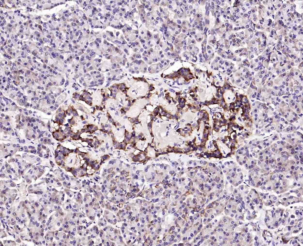
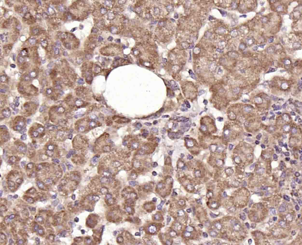
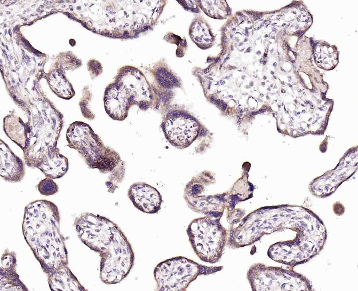
Application
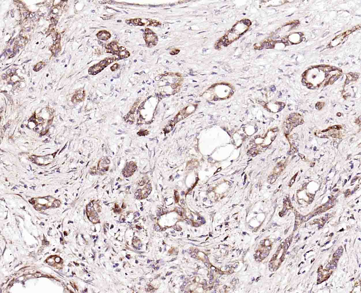
| IHC-P, IHC-F, IF |
|---|---|
| Primary Accession | Q8R3U1 |
| Reactivity | Mouse |
| Host | Rabbit |
| Clonality | Polyclonal |
| Calculated MW | 18 KDa |
| Physical State | Liquid |
| Immunogen | KLH conjugated synthetic peptide derived from the middle of mouse Hrasls3 |
| Epitope Specificity | 8-100/162 |
| Isotype | IgG |
| Purity | affinity purified by Protein A |
| Buffer | 0.01M TBS (pH7.4) with 1% BSA, 0.02% Proclin300 and 50% Glycerol. |
| SUBCELLULAR LOCATION | Membrane; Single-pass membrane protein (Potential). Cytoplasm. Cytoplasm, perinuclear region. |
| SIMILARITY | Belongs to the H-rev107 family. |
| SUBUNIT | Interacts with PPP2R1A; this interaction might decrease PP2A activity. |
| Important Note | This product as supplied is intended for research use only, not for use in human, therapeutic or diagnostic applications. |
| Background Descriptions | HRASLS3 specifically catalyzes the release of fatty acids from phospholipids in adipose tissue and also has a weak lysophospholipase activity. It is a tumor suppressor that may be involved in interferon-dependent cell death. |
| Gene ID | 225845 |
|---|---|
| Other Names | Phospholipase A and acyltransferase 3, Plaat3 {ECO:0000250|UniProtKB:P53816} |
| Target/Specificity | Ubiquitously expressed in normal tissues but down-regulated in primary carcinomas or in many cell lines derived from tumors. Highly expressed in white adipose tissue and in adipocytes. Expressed at lower levels in brown adipose tissue. |
| Dilution | IHC-P=1:100-500,IHC-F=1:100-500,IF=1:100-500 |
| Format | 0.01M TBS(pH7.4), 0.09% (W/V) sodium azide and 50% Glyce |
| Storage | Store at -20 ℃ for one year. Avoid repeated freeze/thaw cycles. When reconstituted in sterile pH 7.4 0.01M PBS or diluent of antibody the antibody is stable for at least two weeks at 2-4 ℃. |
| Name | Plaat3 {ECO:0000250|UniProtKB:P53816} |
|---|---|
| Function | Exhibits both phospholipase A1/2 and acyltransferase activities (PubMed:19047760, PubMed:37919452). Shows phospholipase A1 (PLA1) and A2 (PLA2), catalyzing the calcium-independent release of fatty acids from the sn-1 or sn-2 position of glycerophospholipids (PubMed:18614531, PubMed:19047760, PubMed:19136964, PubMed:22134920). For most substrates, PLA1 activity is much higher than PLA2 activity (By similarity). Shows O-acyltransferase activity, catalyzing the transfer of a fatty acyl group from glycerophospholipid to the hydroxyl group of lysophospholipid (By similarity). Shows N-acyltransferase activity,catalyzing the calcium-independent transfer of a fatty acyl group at the sn-1 position of phosphatidylcholine (PC) and other glycerophospholipids to the primary amine of phosphatidylethanolamine (PE), forming N-acylphosphatidylethanolamine (NAPE), which serves as precursor for N-acylethanolamines (NAEs) (PubMed:19047760). Exhibits high N-acyltransferase activity and low phospholipase A1/2 activity (By similarity). Required for complete organelle rupture and degradation that occur during eye lens terminal differentiation, when fiber cells that compose the lens degrade all membrane-bound organelles in order to provide lens with transparency to allow the passage of light (PubMed:33854238). Organelle membrane degradation is probably catalyzed by the phospholipase activity (PubMed:33854238). |
| Cellular Location | Cell membrane {ECO:0000250|UniProtKB:P53817}; Single-pass membrane protein. Cytoplasm. Cytoplasm, cytosol. Cytoplasm, perinuclear region. Peroxisome membrane; Single-pass membrane protein. Mitochondrion membrane; Single-pass membrane protein. Nucleus envelope. Lysosome membrane; Single-pass membrane protein. Endoplasmic reticulum membrane; Single-pass membrane protein. Note=During eye lens differentiation, recruited from the cytosol to various organelles, including mitochondria, endoplasmic reticulum, nuclear envelope and lysosomes, immediately before organelle degradation. This translocation is triggered by organelle membrane damage and requires the C-terminal transmembrane domain |
| Tissue Location | Ubiquitously expressed in normal tissues but down- regulated in primary carcinomas or in many cell lines derived from tumors (PubMed:12055182). Highly expressed in white adipose tissue and in adipocytes (PubMed:18614531, PubMed:19136964). Expressed at lower levels in brown adipose tissue (PubMed:18614531, PubMed:19136964) |

Thousands of laboratories across the world have published research that depended on the performance of antibodies from Abcepta to advance their research. Check out links to articles that cite our products in major peer-reviewed journals, organized by research category.
info@abcepta.com, and receive a free "I Love Antibodies" mug.

Provided below are standard protocols that you may find useful for product applications.
Background
This product as supplied is intended for research use only, not for use in human, therapeutic or diagnostic applications.
If you have used an Abcepta product and would like to share how it has performed, please click on the "Submit Review" button and provide the requested information. Our staff will examine and post your review and contact you if needed.
If you have any additional inquiries please email technical services at tech@abcepta.com.
Ordering Information
Other Products
Shipping Information
