


 Foundational characteristics of cancer include proliferation, angiogenesis, migration, evasion of apoptosis, and cellular immortality. Find key markers for these cellular processes and antibodies to detect them.
 The SUMOplot™ Analysis Program predicts and scores sumoylation sites in your protein. SUMOylation is a post-translational modification involved in various cellular processes, such as nuclear-cytosolic transport, transcriptional regulation, apoptosis, protein stability, response to stress, and progression through the cell cycle.
 The Autophagy Receptor Motif Plotter predicts and scores autophagy receptor binding sites in your protein. Identifying proteins connected to this pathway is critical to understanding the role of autophagy in physiological as well as pathological processes such as development, differentiation, neurodegenerative diseases, stress, infection, and cancer.
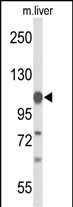
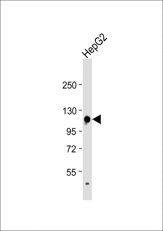
GLDC Antibody (N-term)
Affinity Purified Rabbit Polyclonal Antibody (Pab)
- SPECIFICATION
- CITATIONS: 1
- PROTOCOLS
- BACKGROUND

Application
| WB, E |
|---|---|
| Primary Accession | P23378 |
| Reactivity | Human, Mouse |
| Host | Rabbit |
| Clonality | Polyclonal |
| Isotype | Rabbit IgG |
| Calculated MW | 112730 Da |
| Antigen Region | 49-77 aa |
| Gene ID | 2731 |
|---|---|
| Other Names | Glycine dehydrogenase (decarboxylating), mitochondrial, Glycine cleavage system P protein, Glycine decarboxylase, Glycine dehydrogenase (aminomethyl-transferring), GLDC, GCSP |
| Target/Specificity | This GLDC antibody is generated from rabbits immunized with a KLH conjugated synthetic peptide between 49-77 amino acids from the N-terminal region of human GLDC. |
| Dilution | WB~~1:1000 E~~Use at an assay dependent concentration. |
| Format | Purified polyclonal antibody supplied in PBS with 0.09% (W/V) sodium azide. This antibody is purified through a protein A column, followed by peptide affinity purification. |
| Storage | Maintain refrigerated at 2-8°C for up to 2 weeks. For long term storage store at -20°C in small aliquots to prevent freeze-thaw cycles. |
| Precautions | GLDC Antibody (N-term) is for research use only and not for use in diagnostic or therapeutic procedures. |
| Name | GLDC (HGNC:4313) |
|---|---|
| Function | The glycine cleavage system catalyzes the degradation of glycine. The P protein (GLDC) binds the alpha-amino group of glycine through its pyridoxal phosphate cofactor; CO(2) is released and the remaining methylamine moiety is then transferred to the lipoamide cofactor of the H protein (GCSH). |
| Cellular Location | Mitochondrion. |

Provided below are standard protocols that you may find useful for product applications.
Background
Degradation of glycine is brought about by the glycine cleavage system, which is composed of four mitochondrial protein components: P protein (a pyridoxal phosphate-dependent glycine decarboxylase), H protein (a lipoic acid-containing protein), T protein (a tetrahydrofolate-requiring enzyme), and L protein (a lipoamide dehydrogenase). The protein is the P protein, which binds to glycine and enables the methylamine group from glycine to be transferred to the T protein. Defects in this gene are a cause of nonketotic hyperglycinemia (NKH).
References
Chang, C.Y., et al. Acta Paediatr Taiwan 49(1):35-37(2008)
Kanno, J., et al. J. Med. Genet. 44 (3), E69 (2007)
Kure, S., et al. Hum. Mutat. 27(4):343-352(2006)
If you have used an Abcepta product and would like to share how it has performed, please click on the "Submit Review" button and provide the requested information. Our staff will examine and post your review and contact you if needed.
If you have any additional inquiries please email technical services at tech@abcepta.com.
Ordering Information
Other Products
Shipping Information
