


 Foundational characteristics of cancer include proliferation, angiogenesis, migration, evasion of apoptosis, and cellular immortality. Find key markers for these cellular processes and antibodies to detect them.
 The SUMOplot™ Analysis Program predicts and scores sumoylation sites in your protein. SUMOylation is a post-translational modification involved in various cellular processes, such as nuclear-cytosolic transport, transcriptional regulation, apoptosis, protein stability, response to stress, and progression through the cell cycle.
 The Autophagy Receptor Motif Plotter predicts and scores autophagy receptor binding sites in your protein. Identifying proteins connected to this pathway is critical to understanding the role of autophagy in physiological as well as pathological processes such as development, differentiation, neurodegenerative diseases, stress, infection, and cancer.
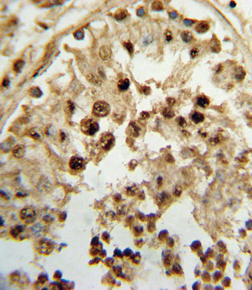
HSD17B3 Antibody (Center)
Affinity Purified Rabbit Polyclonal Antibody (Pab)


- SPECIFICATION
- CITATIONS
- PROTOCOLS
- BACKGROUND

Application
| FC, IHC-P, WB, E |
|---|---|
| Primary Accession | P37058 |
| Reactivity | Human |
| Host | Rabbit |
| Clonality | Polyclonal |
| Isotype | Rabbit IgG |
| Calculated MW | 34516 Da |
| Antigen Region | 89-118 aa |
| Gene ID | 3293 |
|---|---|
| Other Names | Testosterone 17-beta-dehydrogenase 3, 17-beta-hydroxysteroid dehydrogenase type 3, 17-beta-HSD 3, Testicular 17-beta-hydroxysteroid dehydrogenase, HSD17B3, EDH17B3 |
| Target/Specificity | This HSD17B3 antibody is generated from rabbits immunized with a KLH conjugated synthetic peptide between 89-118 amino acids from the Central region of human HSD17B3. |
| Dilution | FC~~1:10~50 IHC-P~~1:10~50 WB~~1:1000 E~~Use at an assay dependent concentration. |
| Format | Purified polyclonal antibody supplied in PBS with 0.09% (W/V) sodium azide. This antibody is purified through a protein A column, followed by peptide affinity purification. |
| Storage | Maintain refrigerated at 2-8°C for up to 2 weeks. For long term storage store at -20°C in small aliquots to prevent freeze-thaw cycles. |
| Precautions | HSD17B3 Antibody (Center) is for research use only and not for use in diagnostic or therapeutic procedures. |
| Name | HSD17B3 (HGNC:5212) |
|---|---|
| Synonyms | EDH17B3, SDR12C2 |
| Function | Catalyzes the conversion of 17-oxosteroids to 17beta- hydroxysteroids (PubMed:16216911, PubMed:26545797, PubMed:27927697, PubMed:8075637). Favors the reduction of androstenedione to testosterone (PubMed:16216911, PubMed:26545797, PubMed:27927697). Testosterone is the key androgen driving male development and function (PubMed:8075637). Uses NADPH while the two other EDH17B enzymes use NADH (PubMed:16216911, PubMed:26545797, PubMed:8075637). Androgens such as epiandrosterone, dehydroepiandrosterone, androsterone and androstanedione are accepted as substrates and reduced at C-17 (PubMed:16216911). Can reduce 11-ketoandrostenedione as well as 11beta- hydroxyandrostenedione at C-17 to the respective testosterone forms (PubMed:16216911, PubMed:27927697). |
| Cellular Location | Endoplasmic reticulum |
| Tissue Location | Testis.. |

Thousands of laboratories across the world have published research that depended on the performance of antibodies from Abcepta to advance their research. Check out links to articles that cite our products in major peer-reviewed journals, organized by research category.
info@abcepta.com, and receive a free "I Love Antibodies" mug.

Provided below are standard protocols that you may find useful for product applications.
Background
This isoform of 17 beta-hydroxysteroid dehydrogenase is expressed predominantly in the testis and catalyzes the conversion of androstenedione to testosterone. It preferentially uses NADP as cofactor. Deficiency can result in male pseudohermaphroditism with gynecomastia.
References
Li, J., et al. Breast Cancer Res. 12 (2), R19 (2010) :
Sata, F., et al. J Sex Med (2010) In press :
Ahn, J., et al. Hum. Mol. Genet. 18(19):3749-3757(2009)
Chakrabarti, B., et al. Autism Res 2(3):157-177(2009)
Beuten, J., et al. Cancer Epidemiol. Biomarkers Prev. 18(6):1869-1880(2009)
Andersson, S., et al. J. Clin. Endocrinol. Metab. 81(1):130-136(1996)
If you have used an Abcepta product and would like to share how it has performed, please click on the "Submit Review" button and provide the requested information. Our staff will examine and post your review and contact you if needed.
If you have any additional inquiries please email technical services at tech@abcepta.com.
Ordering Information
Other Products
Shipping Information
