


 Foundational characteristics of cancer include proliferation, angiogenesis, migration, evasion of apoptosis, and cellular immortality. Find key markers for these cellular processes and antibodies to detect them.
 The SUMOplot™ Analysis Program predicts and scores sumoylation sites in your protein. SUMOylation is a post-translational modification involved in various cellular processes, such as nuclear-cytosolic transport, transcriptional regulation, apoptosis, protein stability, response to stress, and progression through the cell cycle.
 The Autophagy Receptor Motif Plotter predicts and scores autophagy receptor binding sites in your protein. Identifying proteins connected to this pathway is critical to understanding the role of autophagy in physiological as well as pathological processes such as development, differentiation, neurodegenerative diseases, stress, infection, and cancer.
PLA1A Antibody (Center)
Affinity Purified Rabbit Polyclonal Antibody (Pab)
- SPECIFICATION
- CITATIONS
- PROTOCOLS
- BACKGROUND

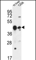
Application
| WB, E |
|---|---|
| Primary Accession | Q53H76 |
| Reactivity | Human, Mouse |
| Host | Rabbit |
| Clonality | Polyclonal |
| Isotype | Rabbit IgG |
| Calculated MW | 49715 Da |
| Antigen Region | 329-358 aa |
| Gene ID | 51365 |
|---|---|
| Other Names | Phospholipase A1 member A, 311-, Phosphatidylserine-specific phospholipase A1, PS-PLA1, PLA1A, NMD, PSPLA1 |
| Target/Specificity | This PLA1A antibody is generated from rabbits immunized with a KLH conjugated synthetic peptide between 329-358 amino acids from the Central region of human PLA1A. |
| Dilution | WB~~1:1000 E~~Use at an assay dependent concentration. |
| Format | Purified polyclonal antibody supplied in PBS with 0.09% (W/V) sodium azide. This antibody is purified through a protein A column, followed by peptide affinity purification. |
| Storage | Maintain refrigerated at 2-8°C for up to 2 weeks. For long term storage store at -20°C in small aliquots to prevent freeze-thaw cycles. |
| Precautions | PLA1A Antibody (Center) is for research use only and not for use in diagnostic or therapeutic procedures. |
| Name | PLA1A (HGNC:17661) |
|---|---|
| Synonyms | NMD, PSPLA1 |
| Function | Hydrolyzes the ester bond of the acyl group attached at the sn-1 position of phosphatidylserines (phospholipase A1 activity) and 1- acyl-2-lysophosphatidylserines (lysophospholipase activity) in the pathway of phosphatidylserines acyl chain remodeling (PubMed:10196188). Cleaves phosphatidylserines exposed on the outer leaflet of the plasma membrane of apoptotic cells producing 2-acyl-1-lysophosphatidylserines, which in turn enhance mast cell activation and histamine production (By similarity). Has no activity toward other glycerophospholipids including phosphatidylcholines, phosphatidylethanolamines, phosphatidic acids or phosphatidylinositols, or glycerolipids such as triolein (By similarity). |
| Cellular Location | Secreted {ECO:0000250|UniProtKB:P97535}. |
| Tissue Location | Widely expressed. Expressed in placenta, prostate and liver. Weakly or not expressed in skin, leukocytes, platelets, colon, spleen, lung, muscle and kidney. |

Thousands of laboratories across the world have published research that depended on the performance of antibodies from Abcepta to advance their research. Check out links to articles that cite our products in major peer-reviewed journals, organized by research category.
info@abcepta.com, and receive a free "I Love Antibodies" mug.

Provided below are standard protocols that you may find useful for product applications.
Background
Phosphatidylserine-specific phospholipase A1-alpha (PLA1A) acts specifically on phosphatidylserine (PS) and 1-acyl-2-lysophosphatidylserine (lyso-PS) to hydrolyze fatty acids at the sn-1 position of these phospholipids.
References
?Morikawa, R.K., et al. J. Biol. Chem. 284(39):26620-26630(2009)
?Aoki, J., et al. Biochim. Biophys. Acta 1582 (1-3), 26-32 (2002) :
?Wang, J., et al. J. Hum. Genet. 47(11):611-613(2002)
?Nagai, Y., et al. J. Biol. Chem. 274(16):11053-11059(1999)
?van Groningen, J.J., et al. FEBS Lett. 404(1):82-86(1997)
If you have used an Abcepta product and would like to share how it has performed, please click on the "Submit Review" button and provide the requested information. Our staff will examine and post your review and contact you if needed.
If you have any additional inquiries please email technical services at tech@abcepta.com.
Ordering Information
Other Products
Shipping Information
