


 Foundational characteristics of cancer include proliferation, angiogenesis, migration, evasion of apoptosis, and cellular immortality. Find key markers for these cellular processes and antibodies to detect them.
 The SUMOplot™ Analysis Program predicts and scores sumoylation sites in your protein. SUMOylation is a post-translational modification involved in various cellular processes, such as nuclear-cytosolic transport, transcriptional regulation, apoptosis, protein stability, response to stress, and progression through the cell cycle.
 The Autophagy Receptor Motif Plotter predicts and scores autophagy receptor binding sites in your protein. Identifying proteins connected to this pathway is critical to understanding the role of autophagy in physiological as well as pathological processes such as development, differentiation, neurodegenerative diseases, stress, infection, and cancer.
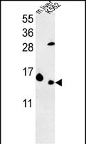
RPL37 Antibody (C-term)
Purified Rabbit Polyclonal Antibody (Pab)
- SPECIFICATION
- CITATIONS: 1
- PROTOCOLS
- BACKGROUND

Application
| WB, E |
|---|---|
| Primary Accession | P61927 |
| Other Accession | P61928, Q9D823, P79244, U3KPD5 |
| Reactivity | Human |
| Predicted | Bovine, Mouse, Rabbit, Rat |
| Host | Rabbit |
| Clonality | Polyclonal |
| Isotype | Rabbit IgG |
| Calculated MW | 11078 Da |
| Antigen Region | 60-88 aa |
| Gene ID | 6167 |
|---|---|
| Other Names | 60S ribosomal protein L37, G116, RPL37 |
| Target/Specificity | This RPL37 antibody is generated from rabbits immunized with a KLH conjugated synthetic peptide between 60-88 amino acids from the C-terminal region of human RPL37. |
| Dilution | WB~~1:1000 E~~Use at an assay dependent concentration. |
| Format | Purified polyclonal antibody supplied in PBS with 0.09% (W/V) sodium azide. This antibody is purified through a protein A column, followed by peptide affinity purification. |
| Storage | Maintain refrigerated at 2-8°C for up to 2 weeks. For long term storage store at -20°C in small aliquots to prevent freeze-thaw cycles. |
| Precautions | RPL37 Antibody (C-term) is for research use only and not for use in diagnostic or therapeutic procedures. |
| Name | RPL37 |
|---|---|
| Function | Component of the large ribosomal subunit (PubMed:23636399, PubMed:32669547). The ribosome is a large ribonucleoprotein complex responsible for the synthesis of proteins in the cell (PubMed:23636399, PubMed:32669547). |
| Cellular Location | Cytoplasm. |

Provided below are standard protocols that you may find useful for product applications.
Background
Ribosomes, the organelles that catalyze protein synthesis, consist of a small 40S subunit and a large 60S subunit. Together these subunits are composed of 4 RNA species and approximately 80 structurally distinct proteins. This gene encodes a ribosomal protein that is a component of the 60S subunit. The protein belongs to the L37E family of ribosomal proteins. It is located in the cytoplasm. The protein contains a C2C2-type zinc finger-like motif.
References
?Ewing, R.M., et al. Mol. Syst. Biol. 3, 89 (2007) :
?Schwartz, E.I., et al. Mol. Cell. Biol. 24(21):9580-9591(2004)
?Kapp, L.D., et al. Annu. Rev. Biochem. 73, 657-704 (2004) :
?Mazumder, B., et al. Cell 115(2):187-198(2003)
?Yoshihama, M., et al. Genome Res. 12(3):379-390(2002)
?Uechi, T., et al. Genomics 72(3):223-230(2001)
?Kenmochi, N., et al. Genome Res. 8(5):509-523(1998)
If you have used an Abcepta product and would like to share how it has performed, please click on the "Submit Review" button and provide the requested information. Our staff will examine and post your review and contact you if needed.
If you have any additional inquiries please email technical services at tech@abcepta.com.
Ordering Information
Shipping Information
