


 Foundational characteristics of cancer include proliferation, angiogenesis, migration, evasion of apoptosis, and cellular immortality. Find key markers for these cellular processes and antibodies to detect them.
 The SUMOplot™ Analysis Program predicts and scores sumoylation sites in your protein. SUMOylation is a post-translational modification involved in various cellular processes, such as nuclear-cytosolic transport, transcriptional regulation, apoptosis, protein stability, response to stress, and progression through the cell cycle.
 The Autophagy Receptor Motif Plotter predicts and scores autophagy receptor binding sites in your protein. Identifying proteins connected to this pathway is critical to understanding the role of autophagy in physiological as well as pathological processes such as development, differentiation, neurodegenerative diseases, stress, infection, and cancer.
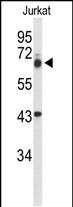
TLE1 Antibody (Center)
Affinity Purified Rabbit Polyclonal Antibody (Pab)
- SPECIFICATION
- CITATIONS
- PROTOCOLS
- BACKGROUND

Application
| WB, E |
|---|---|
| Primary Accession | Q04724 |
| Reactivity | Human |
| Host | Rabbit |
| Clonality | Polyclonal |
| Isotype | Rabbit IgG |
| Calculated MW | 83201 Da |
| Antigen Region | 199-228 aa |
| Gene ID | 7088 |
|---|---|
| Other Names | Transducin-like enhancer protein 1, E(Sp1) homolog, Enhancer of split groucho-like protein 1, ESG1, TLE1 |
| Target/Specificity | This TLE1 antibody is generated from rabbits immunized with a KLH conjugated synthetic peptide between 199-228 amino acids from the Central region of human TLE1. |
| Dilution | WB~~1:1000 E~~Use at an assay dependent concentration. |
| Format | Purified polyclonal antibody supplied in PBS with 0.09% (W/V) sodium azide. This antibody is purified through a protein A column, followed by peptide affinity purification. |
| Storage | Maintain refrigerated at 2-8°C for up to 2 weeks. For long term storage store at -20°C in small aliquots to prevent freeze-thaw cycles. |
| Precautions | TLE1 Antibody (Center) is for research use only and not for use in diagnostic or therapeutic procedures. |
| Name | TLE1 |
|---|---|
| Function | Transcriptional corepressor that binds to a number of transcription factors. Inhibits NF-kappa-B-regulated gene expression. Inhibits the transcriptional activation mediated by FOXA2, and by CTNNB1 and TCF family members in Wnt signaling. Enhances FOXG1/BF- 1- and HES1-mediated transcriptional repression (By similarity). The effects of full-length TLE family members may be modulated by association with dominant-negative AES. Unusual function as coactivator for ESRRG. |
| Cellular Location | Nucleus. Note=Nuclear and chromatin-associated, depending on isoforms and phosphorylation status. Hyperphosphorylation decreases the affinity for nuclear components |
| Tissue Location | In all tissues examined, mostly in brain, liver and muscle |

Thousands of laboratories across the world have published research that depended on the performance of antibodies from Abcepta to advance their research. Check out links to articles that cite our products in major peer-reviewed journals, organized by research category.
info@abcepta.com, and receive a free "I Love Antibodies" mug.

Provided below are standard protocols that you may find useful for product applications.
Background
Transcriptional corepressor that binds to a number of transcription factors. Inhibits NF-kappa-B-regulated gene expression. Inhibits the transcriptional activation mediated by FOXA2, and by CTNNB1 and TCF family members in Wnt signaling. The effects of full-length TLE family members may be modulated by association with dominant-negative AES. Unusual function as coactivator for ESRRG.
References
Ali, S.A., et al. Proc. Natl. Acad. Sci. U.S.A. 107(9):4165-4169(2010)
Jagdis, A., et al. Am. J. Surg. Pathol. 33(12):1743-1751(2009)
Kosemehmetoglu, K., et al. Mod. Pathol. 22(7):872-878(2009)
If you have used an Abcepta product and would like to share how it has performed, please click on the "Submit Review" button and provide the requested information. Our staff will examine and post your review and contact you if needed.
If you have any additional inquiries please email technical services at tech@abcepta.com.
Ordering Information
Other Products
Shipping Information
