


 Foundational characteristics of cancer include proliferation, angiogenesis, migration, evasion of apoptosis, and cellular immortality. Find key markers for these cellular processes and antibodies to detect them.
 The SUMOplot™ Analysis Program predicts and scores sumoylation sites in your protein. SUMOylation is a post-translational modification involved in various cellular processes, such as nuclear-cytosolic transport, transcriptional regulation, apoptosis, protein stability, response to stress, and progression through the cell cycle.
 The Autophagy Receptor Motif Plotter predicts and scores autophagy receptor binding sites in your protein. Identifying proteins connected to this pathway is critical to understanding the role of autophagy in physiological as well as pathological processes such as development, differentiation, neurodegenerative diseases, stress, infection, and cancer.
KRT1 Antibody (Center)
Purified Rabbit Polyclonal Antibody (Pab)
- SPECIFICATION
- CITATIONS: 1
- PROTOCOLS
- BACKGROUND

Application
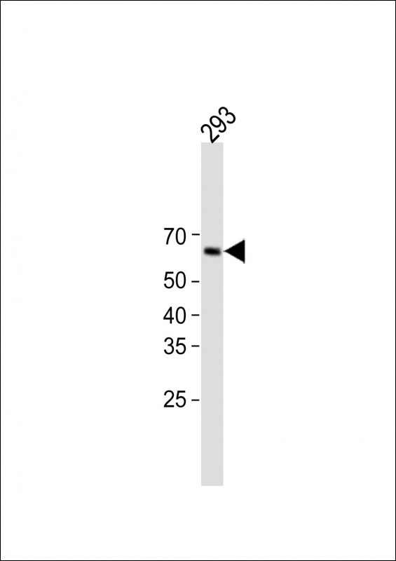
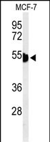
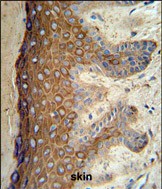
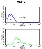
| WB, FC, IHC-P, E |
|---|---|
| Primary Accession | P04264 |
| Reactivity | Human |
| Host | Rabbit |
| Clonality | Polyclonal |
| Isotype | Rabbit IgG |
| Calculated MW | 66039 Da |
| Antigen Region | 415-443 aa |
| Gene ID | 3848 |
|---|---|
| Other Names | Keratin, type II cytoskeletal 1, 67 kDa cytokeratin, Cytokeratin-1, CK-1, Hair alpha protein, Keratin-1, K1, Type-II keratin Kb1, KRT1, KRTA |
| Target/Specificity | This KRT1 antibody is generated from rabbits immunized with a KLH conjugated synthetic peptide between 415-443 amino acids from the Central region of human KRT1. |
| Dilution | WB~~1:1000 FC~~1:10~50 IHC-P~~1:50~100 E~~Use at an assay dependent concentration. |
| Format | Purified polyclonal antibody supplied in PBS with 0.09% (W/V) sodium azide. This antibody is prepared by Saturated Ammonium Sulfate (SAS) precipitation followed by dialysis against PBS. |
| Storage | Maintain refrigerated at 2-8°C for up to 2 weeks. For long term storage store at -20°C in small aliquots to prevent freeze-thaw cycles. |
| Precautions | KRT1 Antibody (Center) is for research use only and not for use in diagnostic or therapeutic procedures. |
| Name | KRT1 |
|---|---|
| Synonyms | KRTA |
| Function | May regulate the activity of kinases such as PKC and SRC via binding to integrin beta-1 (ITB1) and the receptor of activated protein C kinase 1 (RACK1). In complex with C1QBP is a high affinity receptor for kininogen-1/HMWK. |
| Cellular Location | Cell membrane. Cytoplasm |
| Tissue Location | The source of this protein is neonatal foreskin. The 67-kDa type II keratins are expressed in terminally differentiating epidermis |

Provided below are standard protocols that you may find useful for product applications.
Background
KRT1 is a member of the keratin gene family. The type II cytokeratins consist of basic or neutral proteins which are arranged in pairs of heterotypic keratin chains coexpressed during differentiation of simple and stratified epithelial tissues. This type II cytokeratin is specifically expressed in the spinous and granular layers of the epidermis with family member KRT10 and mutations in these genes have been associated with bullous congenital ichthyosiform erythroderma.
References
Labudova, M., et al. J. Virol. 83(16):7842-7849(2009)
Barcelos, A.C., et al. J. Cutan. Pathol. 36(6):647-654(2009)
Grimberg, G., et al. Br. J. Dermatol. 160(2):446-449(2009)
If you have used an Abcepta product and would like to share how it has performed, please click on the "Submit Review" button and provide the requested information. Our staff will examine and post your review and contact you if needed.
If you have any additional inquiries please email technical services at tech@abcepta.com.
Ordering Information
Other Products
Shipping Information
