


 Foundational characteristics of cancer include proliferation, angiogenesis, migration, evasion of apoptosis, and cellular immortality. Find key markers for these cellular processes and antibodies to detect them.
 The SUMOplot™ Analysis Program predicts and scores sumoylation sites in your protein. SUMOylation is a post-translational modification involved in various cellular processes, such as nuclear-cytosolic transport, transcriptional regulation, apoptosis, protein stability, response to stress, and progression through the cell cycle.
 The Autophagy Receptor Motif Plotter predicts and scores autophagy receptor binding sites in your protein. Identifying proteins connected to this pathway is critical to understanding the role of autophagy in physiological as well as pathological processes such as development, differentiation, neurodegenerative diseases, stress, infection, and cancer.
> home > Products > Primary Antibodies > Antibody Collections > SARS CoV2 PPIs > MOSC1 Antibody (Center)
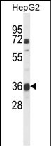
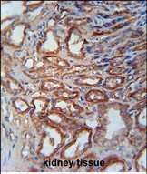
MOSC1 Antibody (Center)
Affinity Purified Rabbit Polyclonal Antibody (Pab)
- SPECIFICATION
- CITATIONS: 3
- PROTOCOLS
- BACKGROUND

Application
| IHC-P, WB, E |
|---|---|
| Primary Accession | Q5VT66 |
| Reactivity | Human |
| Host | Rabbit |
| Clonality | Polyclonal |
| Isotype | Rabbit IgG |
| Calculated MW | 37499 Da |
| Antigen Region | 175-204 aa |
| Gene ID | 64757 |
|---|---|
| Other Names | Mitochondrial amidoxime-reducing component 1, mARC1, 1---, Molybdenum cofactor sulfurase C-terminal domain-containing protein 1, MOSC domain-containing protein 1, Moco sulfurase C-terminal domain-containing protein 1, MARC1, MOSC1 |
| Target/Specificity | This MOSC1 antibody is generated from rabbits immunized with a KLH conjugated synthetic peptide between 175-204 amino acids from the Central region of human MOSC1. |
| Dilution | IHC-P~~1:50~100 WB~~1:1000 E~~Use at an assay dependent concentration. |
| Format | Purified polyclonal antibody supplied in PBS with 0.09% (W/V) sodium azide. This antibody is purified through a protein A column, followed by peptide affinity purification. |
| Storage | Maintain refrigerated at 2-8°C for up to 2 weeks. For long term storage store at -20°C in small aliquots to prevent freeze-thaw cycles. |
| Precautions | MOSC1 Antibody (Center) is for research use only and not for use in diagnostic or therapeutic procedures. |
| Name | MTARC1 (HGNC:26189) |
|---|---|
| Synonyms | MARC1, MOSC1 |
| Function | Catalyzes the reduction of N-oxygenated molecules, acting as a counterpart of cytochrome P450 and flavin-containing monooxygenases in metabolic cycles (PubMed:19053771, PubMed:21029045, PubMed:30397129). As a component of prodrug-converting system, reduces a multitude of N-hydroxylated prodrugs particularly amidoximes, leading to increased drug bioavailability (PubMed:19053771). May be involved in mitochondrial N(omega)-hydroxy-L-arginine (NOHA) reduction, regulating endogenous nitric oxide levels and biosynthesis (PubMed:21029045). Postulated to cleave the N-OH bond of N-hydroxylated substrates in concert with electron transfer from NADH to cytochrome b5 reductase then to cytochrome b5, the ultimate electron donor that primes the active site for substrate reduction (PubMed:19053771, PubMed:21029045). |
| Cellular Location | Mitochondrion outer membrane; Single-pass type II membrane protein. Membrane; Lipid-anchor. Note=Mitochondrial import is mediated by AA 1-40 and requires ATP |
Research Areas
Citations ( 0 )

Application Protocols
Provided below are standard protocols that you may find useful for product applications.
References
Gruenewald, S., et al. J. Med. Chem. 51(24):8173-8177(2008) Anantharaman, V., et al. FEMS Microbiol. Lett. 207(1):55-61(2002)
Abcepta welcomes feedback from its customers.
If you have used an Abcepta product and would like to share how it has performed, please click on the "Submit Review" button and provide the requested information. Our staff will examine and post your review and contact you if needed.
If you have any additional inquiries please email technical services at tech@abcepta.com.
$ 75.00

⚠️ Limit: 5 vials per order for discount.
$ 192.50
⚠️ Limit: 5 vials per order for discount.
Cat# AP9754c
Ordering Information
United States
AlbaniaAustraliaAustriaBelgiumBosnia & HerzegovinaBrazilBulgariaCanadaCentral AmericaChinaCroatiaCyprusCzech RepublicDenmarkEstoniaFinlandFranceGermanyGreeceHong KongHungaryIcelandIndiaIndonesiaIrelandIsraelItalyJapanLatviaLithuaniaLuxembourgMacedoniaMalaysiaMaltaMexicoNetherlandsNew ZealandNorwayPakistanPolandPortugalRomaniaSerbiaSingaporeSlovakiaSloveniaSouth AfricaSouth KoreaSpainSwedenSwitzerlandTaiwanTurkeyUnited KingdomUnited StatesVietnamWorldwideOthers
USA Headquarters
(888) 735-7227 / (858) 622-0099 or (858) 875-1900
Other Products
Shipping Information
Domestic orders (in stock items)
Shipped out the same day. Orders placed after 1 PM (PST) will ship out the next business day.
International orders
Contact your local distributors
