


 Foundational characteristics of cancer include proliferation, angiogenesis, migration, evasion of apoptosis, and cellular immortality. Find key markers for these cellular processes and antibodies to detect them.
 The SUMOplot™ Analysis Program predicts and scores sumoylation sites in your protein. SUMOylation is a post-translational modification involved in various cellular processes, such as nuclear-cytosolic transport, transcriptional regulation, apoptosis, protein stability, response to stress, and progression through the cell cycle.
 The Autophagy Receptor Motif Plotter predicts and scores autophagy receptor binding sites in your protein. Identifying proteins connected to this pathway is critical to understanding the role of autophagy in physiological as well as pathological processes such as development, differentiation, neurodegenerative diseases, stress, infection, and cancer.
MARCO Antibody (N-term)
Affinity Purified Rabbit Polyclonal Antibody (Pab)
- SPECIFICATION
- CITATIONS: 2
- PROTOCOLS
- BACKGROUND

Application
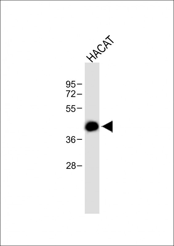
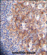
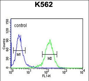
| WB, IHC-P, FC, E |
|---|---|
| Primary Accession | Q9UEW3 |
| Reactivity | Human |
| Host | Rabbit |
| Clonality | Polyclonal |
| Isotype | Rabbit IgG |
| Calculated MW | 52658 Da |
| Antigen Region | 13-40 aa |
| Gene ID | 8685 |
|---|---|
| Other Names | Macrophage receptor MARCO, Macrophage receptor with collagenous structure, Scavenger receptor class A member 2, MARCO, SCARA2 |
| Target/Specificity | This MARCO antibody is generated from rabbits immunized with a KLH conjugated synthetic peptide between 13-40 amino acids of human MARCO. |
| Dilution | WB~~1:500 IHC-P~~1:50~100 FC~~1:10~50 E~~Use at an assay dependent concentration. |
| Format | Purified polyclonal antibody supplied in PBS with 0.09% (W/V) sodium azide. This antibody is purified through a protein A column, followed by peptide affinity purification. |
| Storage | Maintain refrigerated at 2-8°C for up to 2 weeks. For long term storage store at -20°C in small aliquots to prevent freeze-thaw cycles. |
| Precautions | MARCO Antibody (N-term) is for research use only and not for use in diagnostic or therapeutic procedures. |
| Name | MARCO |
|---|---|
| Synonyms | SCARA2 |
| Function | Pattern recognition receptor (PRR) which binds Gram-positive and Gram-negative bacteria (PubMed:9468508). Also plays a role in binding of unopsonized particles by alveolar macrophages (By similarity). Binds to the secretoglobin SCGB3A2 (PubMed:12847263). |
| Cellular Location | Cell membrane; Single-pass type II membrane protein |
| Tissue Location | Expressed in alveolar macrophages (at protein level). Detected in macrophages from various tissues including thymus, kidney, Kupffer cells of liver, and spleen (PubMed:9468508) |

Provided below are standard protocols that you may find useful for product applications.
Background
MARCO is a member of the class A scavenger receptor family and is part of the innate antimicrobial immune system. The protein may bind both Gram-negative and Gram-positive bacteria via an extracellular, C-terminal, scavenger receptor cysteine-rich (SRCR) domain. In addition to short cytoplasmic and transmembrane domains, there is an extracellular spacer domain and a long, extracellular collagenous domain. The protein may form a trimeric molecule by the association of the collagenous domains of three identical polypeptide chains.
References
Wright, A.K., et al. J. Leukoc. Biol. 86(3):479-489(2009)
Trynka, G., et al. Gut 58(8):1078-1083(2009)
Arredouani, M.S., et al. J. Immunol. 175(9):6058-6064(2005)
Liu, T., et al. J. Proteome Res. 4(6):2070-2080(2005)
Seta, N., et al. Arthritis Rheum. 44(4):931-939(2001)
If you have used an Abcepta product and would like to share how it has performed, please click on the "Submit Review" button and provide the requested information. Our staff will examine and post your review and contact you if needed.
If you have any additional inquiries please email technical services at tech@abcepta.com.
Ordering Information
Other Products
Shipping Information
