


 Foundational characteristics of cancer include proliferation, angiogenesis, migration, evasion of apoptosis, and cellular immortality. Find key markers for these cellular processes and antibodies to detect them.
 The SUMOplot™ Analysis Program predicts and scores sumoylation sites in your protein. SUMOylation is a post-translational modification involved in various cellular processes, such as nuclear-cytosolic transport, transcriptional regulation, apoptosis, protein stability, response to stress, and progression through the cell cycle.
 The Autophagy Receptor Motif Plotter predicts and scores autophagy receptor binding sites in your protein. Identifying proteins connected to this pathway is critical to understanding the role of autophagy in physiological as well as pathological processes such as development, differentiation, neurodegenerative diseases, stress, infection, and cancer.
SDR Antibody (Center)
Purified Rabbit Polyclonal Antibody (Pab)
- SPECIFICATION
- CITATIONS: 1
- PROTOCOLS
- BACKGROUND

Application
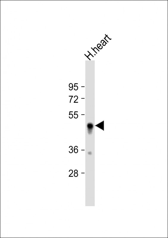
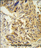
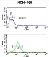
| WB, FC, IHC-P, E |
|---|---|
| Primary Accession | O95810 |
| Reactivity | Human |
| Host | Rabbit |
| Clonality | Polyclonal |
| Isotype | Rabbit IgG |
| Calculated MW | 47173 Da |
| Antigen Region | 109-135 aa |
| Gene ID | 8436 |
|---|---|
| Other Names | Serum deprivation-response protein, Cavin-2, PS-p68, Phosphatidylserine-binding protein, SDPR {ECO:0000312|EMBL:AAD177951} |
| Target/Specificity | This SDR antibody is generated from rabbits immunized with a KLH conjugated synthetic peptide between 109-135 amino acids from the Central region of human SDR. |
| Dilution | WB~~1:1000 FC~~1:10~50 IHC-P~~1:50~100 E~~Use at an assay dependent concentration. |
| Format | Purified polyclonal antibody supplied in PBS with 0.09% (W/V) sodium azide. This antibody is prepared by Saturated Ammonium Sulfate (SAS) precipitation followed by dialysis against PBS. |
| Storage | Maintain refrigerated at 2-8°C for up to 2 weeks. For long term storage store at -20°C in small aliquots to prevent freeze-thaw cycles. |
| Precautions | SDR Antibody (Center) is for research use only and not for use in diagnostic or therapeutic procedures. |
| Name | CAVIN2 (HGNC:10690) |
|---|---|
| Function | Plays an important role in caveolar biogenesis and morphology. Regulates caveolae morphology by inducing membrane curvature within caveolae (PubMed:19525939). Plays a role in caveola formation in a tissue-specific manner. Required for the formation of caveolae in the lung and fat endothelia but not in the heart endothelia. Negatively regulates the size or stability of CAVIN complexes in the lung endothelial cells. May play a role in targeting PRKCA to caveolae (By similarity). |
| Cellular Location | Cytoplasm, cytosol. Membrane, caveola Note=Localizes in the caveolae in a caveolin-dependent manner |
| Tissue Location | Highly expressed in heart and lung, and expressed at lower levels in brain, kidney, liver, pancreas, placenta, and skeletal muscle. |

Provided below are standard protocols that you may find useful for product applications.
Background
This gene encodes a calcium-independent phospholipid-binding protein whose expression increases in serum-starved cells. This protein is a substrate for protein kinase C (PKC) phosphorylation and recruits polymerase I and transcript release factor (PTRF) to caveolae. Removal of this protein causes caveolae loss and its over-expression results in caveolae deformation and membrane tubulation.
References
Baig, A., et al. Proteomics 9(17):4254-4258(2009)
Hansen, C.G., et al. Nat. Cell Biol. 11(7):807-814(2009)
Ogata, T., et al. Mol. Cell. Biol. 28(10):3424-3436(2008)
If you have used an Abcepta product and would like to share how it has performed, please click on the "Submit Review" button and provide the requested information. Our staff will examine and post your review and contact you if needed.
If you have any additional inquiries please email technical services at tech@abcepta.com.

Ordering Information
Other Products
Shipping Information
