


 Foundational characteristics of cancer include proliferation, angiogenesis, migration, evasion of apoptosis, and cellular immortality. Find key markers for these cellular processes and antibodies to detect them.
 The SUMOplot™ Analysis Program predicts and scores sumoylation sites in your protein. SUMOylation is a post-translational modification involved in various cellular processes, such as nuclear-cytosolic transport, transcriptional regulation, apoptosis, protein stability, response to stress, and progression through the cell cycle.
 The Autophagy Receptor Motif Plotter predicts and scores autophagy receptor binding sites in your protein. Identifying proteins connected to this pathway is critical to understanding the role of autophagy in physiological as well as pathological processes such as development, differentiation, neurodegenerative diseases, stress, infection, and cancer.
DNAJC6 Antibody (Center)
Affinity Purified Rabbit Polyclonal Antibody (Pab)
- SPECIFICATION
- CITATIONS
- PROTOCOLS
- BACKGROUND

Application
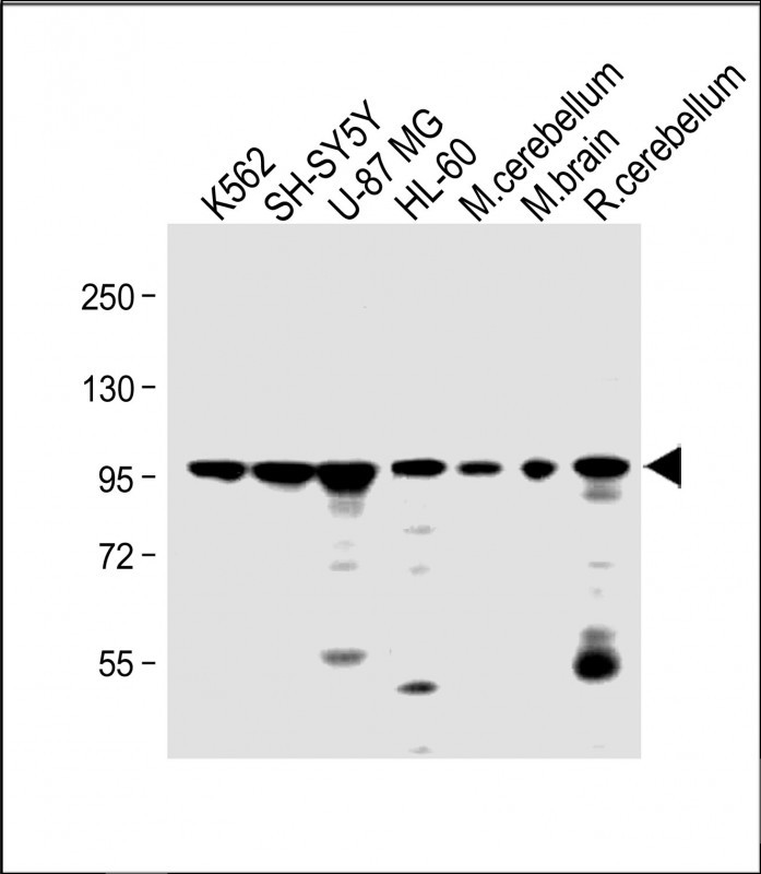
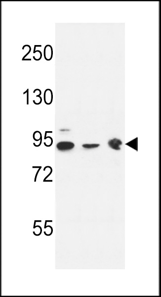
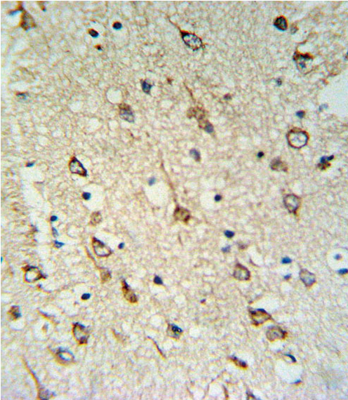
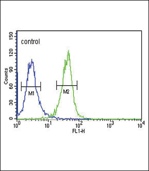
| WB, FC, IHC-P, E |
|---|---|
| Primary Accession | O75061 |
| Other Accession | Q80TZ3 |
| Reactivity | Human, Mouse |
| Host | Rabbit |
| Clonality | Polyclonal |
| Isotype | Rabbit IgG |
| Calculated MW | 99997 Da |
| Antigen Region | 254-281 aa |
| Gene ID | 9829 |
|---|---|
| Other Names | Putative tyrosine-protein phosphatase auxilin, DnaJ homolog subfamily C member 6, DNAJC6, KIAA0473 |
| Target/Specificity | This DNAJC6 antibody is generated from rabbits immunized with a KLH conjugated synthetic peptide between 254-281 amino acids from the Central region of human DNAJC6. |
| Dilution | WB~~1:1000 FC~~1:10~50 IHC-P~~1:10~50 E~~Use at an assay dependent concentration. |
| Format | Purified polyclonal antibody supplied in PBS with 0.09% (W/V) sodium azide. This antibody is purified through a protein A column, followed by peptide affinity purification. |
| Storage | Maintain refrigerated at 2-8°C for up to 2 weeks. For long term storage store at -20°C in small aliquots to prevent freeze-thaw cycles. |
| Precautions | DNAJC6 Antibody (Center) is for research use only and not for use in diagnostic or therapeutic procedures. |
| Name | DNAJC6 (HGNC:15469) |
|---|---|
| Function | May act as a protein phosphatase and/or a lipid phosphatase. Co-chaperone that recruits HSPA8/HSC70 to clathrin-coated vesicles (CCVs) and promotes the ATP-dependent dissociation of clathrin from CCVs and participates in clathrin-mediated endocytosis of synaptic vesicles and their recycling and also in intracellular trafficking (PubMed:18489706). Firstly, binds tightly to the clathrin cages, at a ratio of one DNAJC6 per clathrin triskelion. The HSPA8:ATP complex then binds to the clathrin-auxilin cage, initially at a ratio of one HSPA8 per triskelion leading to ATP hydrolysis stimulation and causing a conformational change in the HSPA8. This cycle is repeated three times to drive to a complex containing the clathrin-auxilin cage associated to three HSPA8:ADP complex. The ATP hydrolysis of the third HSPA8:ATP complex leads to a concerted dismantling of the cage into component triskelia. Then, dissociates from the released triskelia and be recycled to initiate another cycle of HSPA8's recruitment. Also acts during the early steps of clathrin-coated vesicle (CCV) formation through its interaction with the GTP bound form of DNM1 (By similarity). |
| Cellular Location | Cytoplasmic vesicle, clathrin-coated vesicle {ECO:0000250|UniProtKB:Q27974}. Note=Appears on coated vesicles in successive transient bursts, immediately after the vesicle release from the plasma membrane. Recruitment to clathrin-coated vesicles depends on temporal variations in phosphoinositide composition of clathrin-coated vesicles. {ECO:0000250|UniProtKB:Q27974} |
| Tissue Location | Expressed in various brain regions, including cerebellum, corpus callosum, cortex, striatum, brainstem, pons, putamen, spinal cord and substantia nigra. Very low expression in non- neural tissues such as leukocytes, liver, adipose tissue, skeletal muscle and bone marrow. |

Thousands of laboratories across the world have published research that depended on the performance of antibodies from Abcepta to advance their research. Check out links to articles that cite our products in major peer-reviewed journals, organized by research category.
info@abcepta.com, and receive a free "I Love Antibodies" mug.

Provided below are standard protocols that you may find useful for product applications.
Background
DNAJC6 belongs to the evolutionarily conserved DNAJ/HSP40 family of proteins, which regulate molecular chaperone activity by stimulating ATPase activity. DNAJ proteins may have up to 3 distinct domains: a conserved 70-amino acid J domain, usually at the N terminus, a glycine/phenylalanine (G/F)-rich region, and a cysteine-rich domain containing 4 motifs resembling a zinc finger domain
References
Yoshida, T., et al. Int. J. Mol. Med. 25(4):649-656(2010)
Oguri, M., et al. Am. J. Hypertens. 23(1):70-77(2010)
Martins-de-Souza, D., et al. Eur Arch Psychiatry Clin Neurosci 259(3):151-163(2009)
Hirst, J., et al. Traffic 9(8):1354-1371(2008)
If you have used an Abcepta product and would like to share how it has performed, please click on the "Submit Review" button and provide the requested information. Our staff will examine and post your review and contact you if needed.
If you have any additional inquiries please email technical services at tech@abcepta.com.
Ordering Information
Shipping Information
