


 Foundational characteristics of cancer include proliferation, angiogenesis, migration, evasion of apoptosis, and cellular immortality. Find key markers for these cellular processes and antibodies to detect them.
 The SUMOplot™ Analysis Program predicts and scores sumoylation sites in your protein. SUMOylation is a post-translational modification involved in various cellular processes, such as nuclear-cytosolic transport, transcriptional regulation, apoptosis, protein stability, response to stress, and progression through the cell cycle.
 The Autophagy Receptor Motif Plotter predicts and scores autophagy receptor binding sites in your protein. Identifying proteins connected to this pathway is critical to understanding the role of autophagy in physiological as well as pathological processes such as development, differentiation, neurodegenerative diseases, stress, infection, and cancer.
> home > Products > Primary Antibodies > Antibody Collections > Recombinant Antibodies > Anti-TIGIT Reference Antibody (ociperlimab)
Anti-TIGIT Reference Antibody (ociperlimab)
Recombinant Antibody
- SPECIFICATION
- CITATIONS
- PROTOCOLS
- BACKGROUND

Application
| FC, Kinetics, Animal Model |
|---|---|
| Primary Accession | Q495A1 |
| Reactivity | Human |
| Clonality | Monoclonal |
| Isotype | IgG1 |
| Calculated MW | 145.12 KDa |
| Target/Specificity | TIGIT |
|---|---|
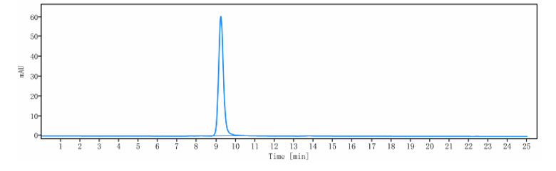
| Endotoxin | < 0.001EU/ µg,determined by LAL method. |
| Conjugation | Unconjugated |
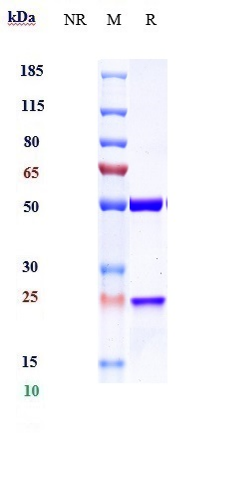
| Expression system | CHO Cell |
| Format | Purified monoclonal antibody supplied in PBS, pH6.0, without preservative.This antibody is purified through a protein A column. |
| Name | TIGIT |
|---|---|
| Synonyms | VSIG9, VSTM3 |
| Function | Inhibitory receptor that plays a role in the modulation of immune responses. Suppresses T-cell activation by promoting the generation of mature immunoregulatory dendritic cells (PubMed:19011627). Upon binding to its ligands PVR/CD155 or NECTIN2/CD112, which are expressed on antigen-presenting cells, sends inhibitory signals to the T-cell or NK cell. Mechanistically, interaction with ligand leads to phosphorylation of the cytoplasmic tail by Src family tyrosine kinases such as FYN or LCK, allowing subsequent binding to adapter GRB2 and SHIP1/INPP5D. In turn, inhibits PI3K and MAPK signaling cascades (PubMed:23154388). In addition, associates with beta-arrestin-2/ARRB2 to recruit SHIP1/INPP5D that suppresses autoubiquitination of TRAF6 and subsequently inhibits NF- kappa-B signaling pathway (PubMed:24817116). Also acts as a receptor for NECTIN4 to inhibit NK cell cytotoxicity (PubMed:32503945). |
| Cellular Location | Cell membrane; Single-pass type I membrane protein. Note=Clustered to the immunological synapse where it disrupts granule polarization and cytotoxicity of NK cells once engaged with PVR. |
| Tissue Location | Expressed at low levels on peripheral memory and regulatory CD4+ T-cells and NK cells and is up-regulated following activation of these cells (at protein level) |
Research Areas
Citations (0)

Thousands of laboratories across the world have published research that depended on the performance of antibodies from Abcepta to advance their research. Check out links to articles that cite our products in major peer-reviewed journals, organized by research category.
Submit your citation using an Abcepta antibody to
info@abcepta.com, and receive a free "I Love Antibodies" mug.
info@abcepta.com, and receive a free "I Love Antibodies" mug.

Application Protocols
Provided below are standard protocols that you may find useful for product applications.
Abcepta welcomes feedback from its customers.
If you have used an Abcepta product and would like to share how it has performed, please click on the "Submit Review" button and provide the requested information. Our staff will examine and post your review and contact you if needed.
If you have any additional inquiries please email technical services at tech@abcepta.com.
$ 385.00
Cat# APR10543
Ordering Information
United States
AlbaniaAustraliaAustriaBelgiumBosnia & HerzegovinaBrazilBulgariaCanadaCentral AmericaChinaCroatiaCyprusCzech RepublicDenmarkEstoniaFinlandFranceGermanyGreeceHong KongHungaryIcelandIndiaIndonesiaIrelandIsraelItalyJapanLatviaLithuaniaLuxembourgMacedoniaMalaysiaMaltaMexicoNetherlandsNew ZealandNorwayPakistanPolandPortugalRomaniaSerbiaSingaporeSlovakiaSloveniaSouth AfricaSouth KoreaSpainSwedenSwitzerlandTaiwanTurkeyUnited KingdomUnited StatesVietnamWorldwideOthers
USA Headquarters
(888) 735-7227 / (858) 622-0099 or (858) 875-1900
Other Products
Shipping Information
Domestic orders (in stock items)
Shipped out the same day. Orders placed after 1 PM (PST) will ship out the next business day.
International orders
Contact your local distributors
