


 Foundational characteristics of cancer include proliferation, angiogenesis, migration, evasion of apoptosis, and cellular immortality. Find key markers for these cellular processes and antibodies to detect them.
 The SUMOplot™ Analysis Program predicts and scores sumoylation sites in your protein. SUMOylation is a post-translational modification involved in various cellular processes, such as nuclear-cytosolic transport, transcriptional regulation, apoptosis, protein stability, response to stress, and progression through the cell cycle.
 The Autophagy Receptor Motif Plotter predicts and scores autophagy receptor binding sites in your protein. Identifying proteins connected to this pathway is critical to understanding the role of autophagy in physiological as well as pathological processes such as development, differentiation, neurodegenerative diseases, stress, infection, and cancer.
> home > Products > Primary Antibodies > Antibody Collections > Recombinant Antibodies > Anti-ENPP3 / CD203c Reference Antibody (Ags-16C3F)
Anti-ENPP3 / CD203c Reference Antibody (Ags-16C3F)
Recombinant Antibody
- SPECIFICATION
- CITATIONS
- PROTOCOLS
- BACKGROUND

Application
| FC, Kinetics, Animal Model |
|---|---|
| Primary Accession | O14638 |
| Reactivity | Human |
| Clonality | Monoclonal |
| Isotype | IgG1 |
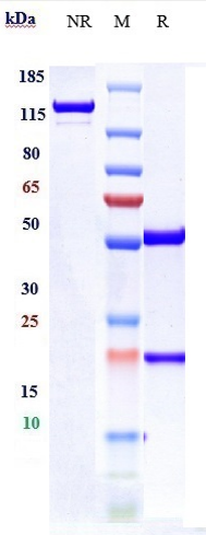
| Calculated MW | 154.04 KDa |
| Target/Specificity | ENPP3 / CD203c |
|---|---|
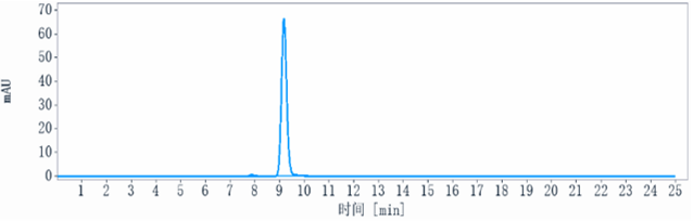
| Endotoxin | < 0.001EU/ µg,determined by LAL method. |
| Conjugation | Unconjugated |
| Expression system | CHO Cell |
| Format | Purified monoclonal antibody supplied in PBS, pH6.0, without preservative.This antibody is purified through a protein A column. |
| Name | ENPP3 (HGNC:3358) |
|---|---|
| Function | Hydrolase that metabolizes extracellular nucleotides, including ATP, GTP, UTP and CTP (PubMed:29717535, PubMed:9344668). Limits mast cells and basophils response during inflammation and during the chronic phases of allergic responses by eliminating extracellular ATP, a signaling molecule activating these cells in an autocrine manner. Metabolizes extracellular ATP in the lumen of the small intestine, and thereby prevents ATP-induced apoptosis of intestinal plasmacytoid dendritic cells (By similarity). Has a broad specificity and can also hydrolyze UDP-GlcNAc into UMP and GlcNAc-1-phosphate and potentially several other intracellular nucleotide sugars, including UDP-GalNAc, CMP-NeuAc, GDP-Fuc, and UDP-GlcA. Thereby, could modulate glycan biosynthesis and protein glycosylation (By similarity). Can hydrolyze extracellular dinucleoside polyphosphates, including the vasoactive adenosine polyphosphates as well (PubMed:12846830). In addition, displays an alkaline phosphodiesterase activity in vitro (PubMed:11342463). |
| Cellular Location | Cell membrane; Single-pass type II membrane protein. Apical cell membrane; Single-pass type II membrane protein. Secreted Note=Detected at the cell surface of basophils (PubMed:11342463) Detected at the apical plasma membrane of bile duct cells (PubMed:15072822). Located to the apical surface in intestinal and kidney epithelial cells. Secreted in serum, and in lumen of epithelial cells. |
| Tissue Location | Detected on bile ducts in liver, and in blood serum (at protein level) (PubMed:15072822). Detected in prostate and uterus (PubMed:9344668). Detected on basophils, but not neutrophils (PubMed:11342463). |
Research Areas
Citations (0)

Thousands of laboratories across the world have published research that depended on the performance of antibodies from Abcepta to advance their research. Check out links to articles that cite our products in major peer-reviewed journals, organized by research category.
Submit your citation using an Abcepta antibody to
info@abcepta.com, and receive a free "I Love Antibodies" mug.
info@abcepta.com, and receive a free "I Love Antibodies" mug.

Application Protocols
Provided below are standard protocols that you may find useful for product applications.
Abcepta welcomes feedback from its customers.
If you have used an Abcepta product and would like to share how it has performed, please click on the "Submit Review" button and provide the requested information. Our staff will examine and post your review and contact you if needed.
If you have any additional inquiries please email technical services at tech@abcepta.com.
$ 385.00
Cat# APR10726
Ordering Information
United States
AlbaniaAustraliaAustriaBelgiumBosnia & HerzegovinaBrazilBulgariaCanadaCentral AmericaChinaCroatiaCyprusCzech RepublicDenmarkEstoniaFinlandFranceGermanyGreeceHong KongHungaryIcelandIndiaIndonesiaIrelandIsraelItalyJapanLatviaLithuaniaLuxembourgMacedoniaMalaysiaMaltaMexicoNetherlandsNew ZealandNorwayPakistanPolandPortugalRomaniaSerbiaSingaporeSlovakiaSloveniaSouth AfricaSouth KoreaSpainSwedenSwitzerlandTaiwanTurkeyUnited KingdomUnited StatesVietnamWorldwideOthers
USA Headquarters
(888) 735-7227 / (858) 622-0099 or (858) 875-1900
Other Products
Shipping Information
Domestic orders (in stock items)
Shipped out the same day. Orders placed after 1 PM (PST) will ship out the next business day.
International orders
Contact your local distributors
