


 Foundational characteristics of cancer include proliferation, angiogenesis, migration, evasion of apoptosis, and cellular immortality. Find key markers for these cellular processes and antibodies to detect them.
 The SUMOplot™ Analysis Program predicts and scores sumoylation sites in your protein. SUMOylation is a post-translational modification involved in various cellular processes, such as nuclear-cytosolic transport, transcriptional regulation, apoptosis, protein stability, response to stress, and progression through the cell cycle.
 The Autophagy Receptor Motif Plotter predicts and scores autophagy receptor binding sites in your protein. Identifying proteins connected to this pathway is critical to understanding the role of autophagy in physiological as well as pathological processes such as development, differentiation, neurodegenerative diseases, stress, infection, and cancer.
> home > Products > Primary Antibodies > Antibody Collections > Recombinant Antibodies > Anti-CEACAM6 / CD66c Reference Antibody (NEO-201)
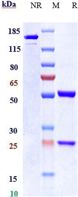
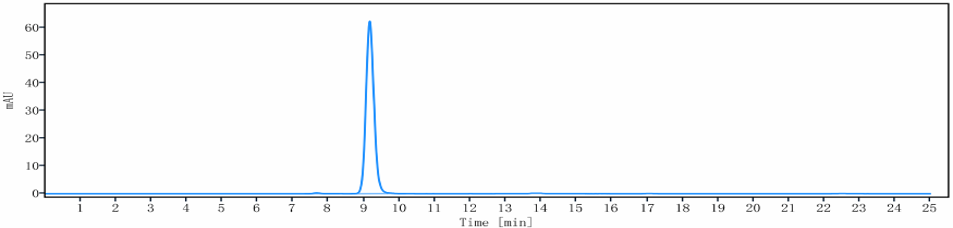
Anti-CEACAM6 / CD66c Reference Antibody (NEO-201)
Recombinant Antibody
- SPECIFICATION
- CITATIONS
- PROTOCOLS
- BACKGROUND

Application
| FC, Kinetics, Animal Model |
|---|---|
| Primary Accession | P40199 |
| Reactivity | Human |
| Clonality | Monoclonal |
| Isotype | IgG1 |
| Calculated MW | 145.48 KDa |
| Target/Specificity | CEACAM6 / CD66c |
|---|---|
| Endotoxin | < 0.001EU/ µg,determined by LAL method. |
| Conjugation | Unconjugated |
| Expression system | CHO Cell |
| Format | Purified monoclonal antibody supplied in PBS, pH6.0, without preservative.This antibody is purified through a protein A column. |
| Name | CEACAM6 (HGNC:1818) |
|---|---|
| Function | Cell surface glycoprotein that plays a role in cell adhesion and tumor progression (PubMed:10910050, PubMed:11590190, PubMed:1378450, PubMed:16204051, PubMed:2022629, PubMed:2803308, PubMed:8776764). Intercellular adhesion occurs in a calcium- and fibronectin-independent manner (PubMed:16204051, PubMed:2022629). Mediates homophilic and heterophilic cell adhesion with other carcinoembryonic antigen-related cell adhesion molecules, such as CEACAM5 and CEACAM8 (PubMed:11590190, PubMed:16204051, PubMed:2022629, PubMed:2803308, PubMed:8776764). Heterophilic interaction with CEACAM8 occurs in activated neutrophils (PubMed:8776764). Plays a role in neutrophil adhesion to cytokine-activated endothelial cells (PubMed:1378450). Plays a role in cell migration and cell adhesion to endothelial cells (PubMed:16204051). |
| Cellular Location | Cell membrane; Lipid-anchor, GPI-anchor. Apical cell membrane. Cell surface. Note=Localized to the apical glycocalyx surface. |
| Tissue Location | Expressed in neutrophils (PubMed:1378450). Expressed in columnar epithelial and goblet cells of the colon (PubMed:10436421). Expressed in numerous tumor cell lines (at protein level) (PubMed:16204051). |
Research Areas
Citations (0)

Thousands of laboratories across the world have published research that depended on the performance of antibodies from Abcepta to advance their research. Check out links to articles that cite our products in major peer-reviewed journals, organized by research category.
Submit your citation using an Abcepta antibody to
info@abcepta.com, and receive a free "I Love Antibodies" mug.
info@abcepta.com, and receive a free "I Love Antibodies" mug.

Application Protocols
Provided below are standard protocols that you may find useful for product applications.
Abcepta welcomes feedback from its customers.
If you have used an Abcepta product and would like to share how it has performed, please click on the "Submit Review" button and provide the requested information. Our staff will examine and post your review and contact you if needed.
If you have any additional inquiries please email technical services at tech@abcepta.com.
$ 385.00
Cat# APR10829
Ordering Information
United States
AlbaniaAustraliaAustriaBelgiumBosnia & HerzegovinaBrazilBulgariaCanadaCentral AmericaChinaCroatiaCyprusCzech RepublicDenmarkEstoniaFinlandFranceGermanyGreeceHong KongHungaryIcelandIndiaIndonesiaIrelandIsraelItalyJapanLatviaLithuaniaLuxembourgMacedoniaMalaysiaMaltaMexicoNetherlandsNew ZealandNorwayPakistanPolandPortugalRomaniaSerbiaSingaporeSlovakiaSloveniaSouth AfricaSouth KoreaSpainSwedenSwitzerlandTaiwanTurkeyUnited KingdomUnited StatesVietnamWorldwideOthers
USA Headquarters
(888) 735-7227 / (858) 622-0099 or (858) 875-1900
Other Products
Shipping Information
Domestic orders (in stock items)
Shipped out the same day. Orders placed after 1 PM (PST) will ship out the next business day.
International orders
Contact your local distributors
