


 Foundational characteristics of cancer include proliferation, angiogenesis, migration, evasion of apoptosis, and cellular immortality. Find key markers for these cellular processes and antibodies to detect them.
 The SUMOplot™ Analysis Program predicts and scores sumoylation sites in your protein. SUMOylation is a post-translational modification involved in various cellular processes, such as nuclear-cytosolic transport, transcriptional regulation, apoptosis, protein stability, response to stress, and progression through the cell cycle.
 The Autophagy Receptor Motif Plotter predicts and scores autophagy receptor binding sites in your protein. Identifying proteins connected to this pathway is critical to understanding the role of autophagy in physiological as well as pathological processes such as development, differentiation, neurodegenerative diseases, stress, infection, and cancer.
> home > Products > Primary Antibodies > Antibody Collections > Recombinant Antibodies > Anti-CLEC7A Reference Antibody (Baylor patent anti-Dectin-1)
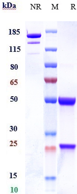
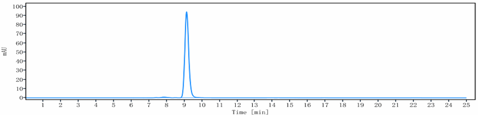
Anti-CLEC7A Reference Antibody (Baylor patent anti-Dectin-1)
Recombinant Antibody
- SPECIFICATION
- CITATIONS
- PROTOCOLS
- BACKGROUND

Application
| FC, Kinetics, Animal Model |
|---|---|
| Primary Accession | Q9BXN2 |
| Reactivity | Human |
| Clonality | Monoclonal |
| Isotype | IgG4 |
| Calculated MW | 150 KDa |
| Target/Specificity | CLEC7A |
|---|---|
| Endotoxin | < 0.001EU/ µg,determined by LAL method. |
| Conjugation | Unconjugated |
| Expression system | CHO Cell |
| Format | Purified monoclonal antibody supplied in PBS, pH6.0, without preservative.This antibody is purified through a protein A column. |
| Name | CLEC7A (HGNC:14558) |
|---|---|
| Function | Lectin that functions as a pattern recognizing receptor (PRR) specific for beta-1,3-linked and beta-1,6-linked glucans, which constitute cell wall constituents from pathogenic bacteria and fungi (PubMed:11567029, PubMed:12423684). Necessary for the TLR2-mediated inflammatory response and activation of NF-kappa-B: upon beta-glucan binding, recruits SYK via its ITAM motif and promotes a signaling cascade that activates some CARD domain-BCL10-MALT1 (CBM) signalosomes, leading to the activation of NF-kappa-B and MAP kinase p38 (MAPK11, MAPK12, MAPK13 and/or MAPK14) pathways which stimulate expression of genes encoding pro-inflammatory cytokines and chemokines (By similarity). Enhances cytokine production in macrophages and dendritic cells (By similarity). Mediates production of reactive oxygen species in the cell (By similarity). Mediates phagocytosis of C.albicans conidia (PubMed:17230442). Binds T-cells in a way that does not involve their surface glycans and plays a role in T-cell activation. Stimulates T-cell proliferation. Induces phosphorylation of SCIMP after binding beta-glucans (By similarity). |
| Cellular Location | Cell membrane; Single-pass type II membrane protein [Isoform 6]: Cytoplasm. |
| Tissue Location | Highly expressed in peripheral blood leukocytes and dendritic cells. Detected in spleen, bone marrow, lung, muscle, stomach and placenta. |
Research Areas
Citations (0)

Thousands of laboratories across the world have published research that depended on the performance of antibodies from Abcepta to advance their research. Check out links to articles that cite our products in major peer-reviewed journals, organized by research category.
Submit your citation using an Abcepta antibody to
info@abcepta.com, and receive a free "I Love Antibodies" mug.
info@abcepta.com, and receive a free "I Love Antibodies" mug.

Application Protocols
Provided below are standard protocols that you may find useful for product applications.
Abcepta welcomes feedback from its customers.
If you have used an Abcepta product and would like to share how it has performed, please click on the "Submit Review" button and provide the requested information. Our staff will examine and post your review and contact you if needed.
If you have any additional inquiries please email technical services at tech@abcepta.com.
$ 385.00
Cat# APR10838
Ordering Information
United States
AlbaniaAustraliaAustriaBelgiumBosnia & HerzegovinaBrazilBulgariaCanadaCentral AmericaChinaCroatiaCyprusCzech RepublicDenmarkEstoniaFinlandFranceGermanyGreeceHong KongHungaryIcelandIndiaIndonesiaIrelandIsraelItalyJapanLatviaLithuaniaLuxembourgMacedoniaMalaysiaMaltaMexicoNetherlandsNew ZealandNorwayPakistanPolandPortugalRomaniaSerbiaSingaporeSlovakiaSloveniaSouth AfricaSouth KoreaSpainSwedenSwitzerlandTaiwanTurkeyUnited KingdomUnited StatesVietnamWorldwideOthers
USA Headquarters
(888) 735-7227 / (858) 622-0099 or (858) 875-1900
Other Products
Shipping Information
Domestic orders (in stock items)
Shipped out the same day. Orders placed after 1 PM (PST) will ship out the next business day.
International orders
Contact your local distributors
