


 Foundational characteristics of cancer include proliferation, angiogenesis, migration, evasion of apoptosis, and cellular immortality. Find key markers for these cellular processes and antibodies to detect them.
 The SUMOplot™ Analysis Program predicts and scores sumoylation sites in your protein. SUMOylation is a post-translational modification involved in various cellular processes, such as nuclear-cytosolic transport, transcriptional regulation, apoptosis, protein stability, response to stress, and progression through the cell cycle.
 The Autophagy Receptor Motif Plotter predicts and scores autophagy receptor binding sites in your protein. Identifying proteins connected to this pathway is critical to understanding the role of autophagy in physiological as well as pathological processes such as development, differentiation, neurodegenerative diseases, stress, infection, and cancer.
> home > Products > Primary Antibodies > Antibody Collections > Recombinant Antibodies > Anti-PMEL Reference Antibody (Genentech anti-PMEL17)
Anti-PMEL Reference Antibody (Genentech anti-PMEL17)
Recombinant Antibody
- SPECIFICATION
- CITATIONS
- PROTOCOLS
- BACKGROUND

Application
| FC, Kinetics, Animal Model |
|---|---|
| Primary Accession | P40967 |
| Reactivity | Rat, Human, Mouse |
| Clonality | Monoclonal |
| Isotype | IgG1 |
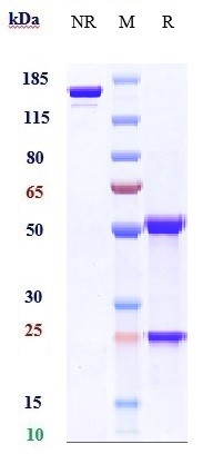
| Calculated MW | 145.54 KDa |
| Target/Specificity | PMEL |
|---|---|
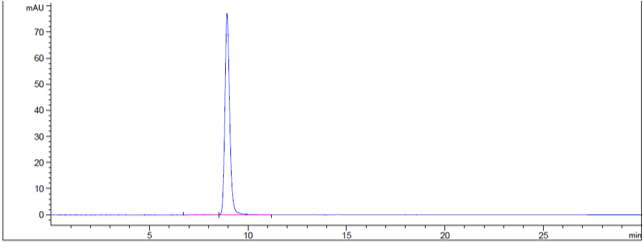
| Endotoxin | < 0.001EU/ µg,determined by LAL method. |
| Conjugation | Unconjugated |
| Expression system | CHO Cell |
| Format | Purified monoclonal antibody supplied in PBS, pH6.0, without preservative.This antibody is purified through a protein A column. |
| Name | PMEL |
|---|---|
| Synonyms | D12S53E, PMEL17, SILV |
| Function | Forms physiological amyloids that play a central role in melanosome morphogenesis and pigmentation. The maturation of unpigmented premelanosomes from stage I to II is marked by assembly of processed amyloidogenic fragments into parallel fibrillar sheets, which elongate the vesicle into a striated ellipsoidal shape. In pigmented stage III and IV melanosomes, the amyloid matrix serves as a platform where eumelanin precursors accumulate at high local concentrations for pigment formation. May prevent pigmentation-associated toxicity by sequestering toxic reaction intermediates of eumelanin biosynthesis pathway. |
| Cellular Location | Endoplasmic reticulum membrane; Single-pass type I membrane protein. Golgi apparatus, cis-Golgi network membrane; Single-pass type I membrane protein. Endosome, multivesicular body. Melanosome Extracellular vesicle. Secreted. Note=Identified by mass spectrometry in melanosome fractions from stage I to stage IV (PubMed:17081065) Localizes predominantly to intralumenal vesicles (ILVs) within multivesicular bodies. Associates with ILVs found within the lumen of premelanosomes and melanosomes and particularly in compartments that serve as precursors to the striated stage II premelanosomes (PubMed:11694580, PubMed:12643545). Sorted to stage I melanosomes following its processing in the ER and cis-Golgi (PubMed:15096515) Transiently expressed at the cell surface before targeting to early melanosomes (PubMed:16760433, PubMed:30988362). Colocalizes with BACE2 in stage I and II melanosomes (PubMed:23754390). Colocalizes with CD63 and APOE at exosomes and in intraluminal vesicles within multivesicular endosomes (PubMed:21962903, PubMed:26387950) |
| Tissue Location | Normally expressed at low levels in quiescent adult melanocytes but overexpressed by proliferating neonatal melanocytes and during tumor growth. Overexpressed in melanomas. Some expression was found in dysplastic nevi. |
Research Areas
Citations (0)

Thousands of laboratories across the world have published research that depended on the performance of antibodies from Abcepta to advance their research. Check out links to articles that cite our products in major peer-reviewed journals, organized by research category.
Submit your citation using an Abcepta antibody to
info@abcepta.com, and receive a free "I Love Antibodies" mug.
info@abcepta.com, and receive a free "I Love Antibodies" mug.

Application Protocols
Provided below are standard protocols that you may find useful for product applications.
Abcepta welcomes feedback from its customers.
If you have used an Abcepta product and would like to share how it has performed, please click on the "Submit Review" button and provide the requested information. Our staff will examine and post your review and contact you if needed.
If you have any additional inquiries please email technical services at tech@abcepta.com.
$ 385.00
Cat# APR11014
Ordering Information
United States
AlbaniaAustraliaAustriaBelgiumBosnia & HerzegovinaBrazilBulgariaCanadaCentral AmericaChinaCroatiaCyprusCzech RepublicDenmarkEstoniaFinlandFranceGermanyGreeceHong KongHungaryIcelandIndiaIndonesiaIrelandIsraelItalyJapanLatviaLithuaniaLuxembourgMacedoniaMalaysiaMaltaMexicoNetherlandsNew ZealandNorwayPakistanPolandPortugalRomaniaSerbiaSingaporeSlovakiaSloveniaSouth AfricaSouth KoreaSpainSwedenSwitzerlandTaiwanTurkeyUnited KingdomUnited StatesVietnamWorldwideOthers
USA Headquarters
(888) 735-7227 / (858) 622-0099 or (858) 875-1900
Other Products
Shipping Information
Domestic orders (in stock items)
Shipped out the same day. Orders placed after 1 PM (PST) will ship out the next business day.
International orders
Contact your local distributors
