


 Foundational characteristics of cancer include proliferation, angiogenesis, migration, evasion of apoptosis, and cellular immortality. Find key markers for these cellular processes and antibodies to detect them.
 The SUMOplot™ Analysis Program predicts and scores sumoylation sites in your protein. SUMOylation is a post-translational modification involved in various cellular processes, such as nuclear-cytosolic transport, transcriptional regulation, apoptosis, protein stability, response to stress, and progression through the cell cycle.
 The Autophagy Receptor Motif Plotter predicts and scores autophagy receptor binding sites in your protein. Identifying proteins connected to this pathway is critical to understanding the role of autophagy in physiological as well as pathological processes such as development, differentiation, neurodegenerative diseases, stress, infection, and cancer.
DcR2 Antibody
- SPECIFICATION
- CITATIONS
- PROTOCOLS
- BACKGROUND

Application
| WB, ICC, E |
|---|---|
| Primary Accession | Q9UBN6 |
| Other Accession | Q9UBN6, 18203495 |
| Reactivity | Human, Mouse, Rat |
| Host | Rabbit |
| Clonality | Polyclonal |
| Isotype | IgG |
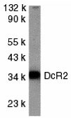
| Calculated MW | Predicted: 36 kDa Observed: 36 kDa |
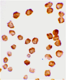
| Application Notes | DcR2 antibody can be used for detection of DcR2 expression by Western blot at 1 µg/mL. Antibody can also be used for immunocytochemistry starting at 10 µg/mL. |
| Gene ID | 8793 |
|---|---|
| Other Names | DcR2 Antibody: DCR2, CD264, TRUNDD, TRAILR4, TRAIL-R4, DCR2, UNQ251/PRO288, Tumor necrosis factor receptor superfamily member 10D, Decoy receptor 2, DcR2, tumor necrosis factor receptor superfamily, member 10d, decoy with truncated death domain |
| Target/Specificity | TNFRSF10D; |
| Reconstitution & Storage | DcR2 antibody can be stored at 4℃ for three months and -20℃, stable for up to one year. As with all antibodies care should be taken to avoid repeated freeze thaw cycles. Antibodies should not be exposed to prolonged high temperatures. |
| Precautions | DcR2 Antibody is for research use only and not for use in diagnostic or therapeutic procedures. |
| Name | TNFRSF10D (HGNC:11907) |
|---|---|
| Function | Receptor for the cytotoxic ligand TRAIL (PubMed:9430226). Contains a truncated death domain and hence is not capable of inducing apoptosis but protects against TRAIL-mediated apoptosis (PubMed:9537512). Reports are contradictory with regards to its ability to induce the NF-kappa-B pathway. According to PubMed:9382840, it cannot but according to PubMed:9430226, it can induce the NF-kappa-B pathway (PubMed:9382840, PubMed:9430226). |
| Cellular Location | Membrane; Single-pass type I membrane protein |
| Tissue Location | Widely expressed, in particular in fetal kidney, lung and liver, and in adult testis and liver. Also expressed in peripheral blood leukocytes, colon and small intestine, ovary, prostate, thymus, spleen, pancreas, kidney, lung, placenta and heart |

Thousands of laboratories across the world have published research that depended on the performance of antibodies from Abcepta to advance their research. Check out links to articles that cite our products in major peer-reviewed journals, organized by research category.
info@abcepta.com, and receive a free "I Love Antibodies" mug.

Provided below are standard protocols that you may find useful for product applications.
Background
DcR2 Antibody: Apoptosis is induced by certain cytokines including TNF and Fas ligand in the TNF family through their death domain containing receptors. TRAIL/Apo2L is a new member of the TNF family and induces apoptosis of a variety of tumor cell lines. DR4 and DR5 are the recently identified functional receptors for TRAIL, and DcR1/TRID is a decoy receptor. Another member of the TRAIL receptor family was more recently identified and designated DcR2, TRAIL-R4, or TRUNDD. DcR2 has an extracellular TRAIL-binding domain but lacks intracellular death domain and does not induce apoptosis. Like DR4 and DR5, DcR2 transcript is widely expressed in normal human tissues. Overexpression of DcR2 attenuated TRAIL-induced apoptosis.
References
Pan G, O'Rourke K, Chinnaiyan AM, et al. The receptor for the cytotoxic ligand TRAIL. Science 1997; 276:111-3.
Pan G, Ni J, Wei YF, Yu G, et al. An antagonist decoy receptor and a death domain-containing receptor for TRAIL. Science 1997; 277:815-8.
Sheridan JP, Marsters SA, Pitti RM, et al. Control of TRAIL-induced apoptosis by a family of signaling and decoy receptors. Science 1997; 277:818-21.
Marsters SA, Sheridan JP, Pitti RM, et al. A novel receptor for Apo2L/TRAIL contains a truncated death domain. Curr. Biol. 1997; 7:1003-6.
If you have used an Abcepta product and would like to share how it has performed, please click on the "Submit Review" button and provide the requested information. Our staff will examine and post your review and contact you if needed.
If you have any additional inquiries please email technical services at tech@abcepta.com.
Ordering Information
Other Products
Shipping Information
