


 Foundational characteristics of cancer include proliferation, angiogenesis, migration, evasion of apoptosis, and cellular immortality. Find key markers for these cellular processes and antibodies to detect them.
 The SUMOplot™ Analysis Program predicts and scores sumoylation sites in your protein. SUMOylation is a post-translational modification involved in various cellular processes, such as nuclear-cytosolic transport, transcriptional regulation, apoptosis, protein stability, response to stress, and progression through the cell cycle.
 The Autophagy Receptor Motif Plotter predicts and scores autophagy receptor binding sites in your protein. Identifying proteins connected to this pathway is critical to understanding the role of autophagy in physiological as well as pathological processes such as development, differentiation, neurodegenerative diseases, stress, infection, and cancer.
TAB1 Antibody
- SPECIFICATION
- CITATIONS
- PROTOCOLS
- BACKGROUND

Application
| WB, IF, ICC, E |
|---|---|
| Primary Accession | Q15750 |
| Other Accession | NP_006107, 5174703 |
| Reactivity | Human, Mouse |
| Host | Rabbit |
| Clonality | Polyclonal |
| Isotype | IgG |
| Calculated MW | 54644 Da |
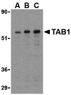
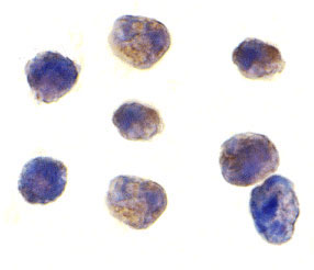
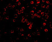
| Application Notes | TAB1 antibody can be used for the detection of TAB1 by Western blot at 0.5 to 2 µg/mL. Antibody can also be used for immunocytochemistry starting at 1 µg/mL. For immunofluorescence start at 2 µg/mL. |
| Gene ID | 10454 |
|---|---|
| Other Names | TAB1 Antibody: 3'-Tab1, MAP3K7IP1, TGF-beta-activated kinase 1 and MAP3K7-binding protein 1, Mitogen-activated protein kinase kinase kinase 7-interacting protein 1, TAK1-binding protein 1, mitogen-activated protein kinase kinase kinase 7 interacting protein 1 |
| Target/Specificity | MAP3K7IP1; |
| Reconstitution & Storage | TAB1 antibody can be stored at 4℃ for three months and -20℃, stable for up to one year. As with all antibodies care should be taken to avoid repeated freeze thaw cycles. Antibodies should not be exposed to prolonged high temperatures. |
| Precautions | TAB1 Antibody is for research use only and not for use in diagnostic or therapeutic procedures. |
| Name | TAB1 |
|---|---|
| Synonyms | MAP3K7IP1 |
| Function | Key adapter protein that plays an essential role in JNK and NF-kappa-B activation and proinflammatory cytokines production in response to stimulation with TLRs and cytokines (PubMed:22307082, PubMed:24403530). Mechanistically, associates with the catalytic domain of MAP3K7/TAK1 to trigger MAP3K7/TAK1 autophosphorylation leading to its full activation (PubMed:10838074, PubMed:25260751, PubMed:37832545). Similarly, associates with MAPK14 and triggers its autophosphorylation and subsequent activation (PubMed:11847341, PubMed:29229647). In turn, MAPK14 phosphorylates TAB1 and inhibits MAP3K7/TAK1 activation in a feedback control mechanism (PubMed:14592977). Also plays a role in recruiting MAPK14 to the TAK1 complex for the phosphorylation of the TAB2 and TAB3 regulatory subunits (PubMed:18021073). |
| Cellular Location | Cytoplasm, cytosol. Endoplasmic reticulum membrane; Peripheral membrane protein; Cytoplasmic side. Note=Recruited to the endoplasmic reticulum following interaction with STING1 |
| Tissue Location | Ubiquitous.. |

Thousands of laboratories across the world have published research that depended on the performance of antibodies from Abcepta to advance their research. Check out links to articles that cite our products in major peer-reviewed journals, organized by research category.
info@abcepta.com, and receive a free "I Love Antibodies" mug.

Provided below are standard protocols that you may find useful for product applications.
Background
TAB1 Antibody: TAB1 was identified as a regulator of the MAP kinase kinase kinase TAK1/MAP3K7, which is known to mediate various intracellular signaling pathways, such as those induced by TGF-beta and members of the Toll-IL-1R (TIR) superfamily, thus acting as an intermediate in both proliferative and innate and adaptive immune responses. This protein, together with either TAB2 or TAB3, activates TAK1 kinase in response to upstream signals. It has been shown that the C-terminal portion of TAB1 is sufficient for binding and activation of TAK1, while a portion of the N-terminus acts as a dominant-negative inhibitor of TGF-beta, demonstrating how this protein can function as a mediator between TGF-beta receptors and TAK1.
References
Shibuya H, Yamaguchi K, Shirakabe K, et al. TAB1: an activator of the TAK1 MAPKKK in TGF-β signal transduction. Science 1996; 272:1179-82.
Irie T, Muta T, and Takeshige K. TAK1 mediates an activation signal from toll-like receptor(s) to nuclear factor-κB in lipopolysaccharide-stimulated macrophages. FEBS Lett. 2000; 467:160-4.
Akira S and Takeda K. Toll-like receptor Signalling. Nat. Rev. Immunol. 2004; 4:499-511.
Jiang Z, Ninomiya-Tsuji J, Qian Y, et al. Interleukin-1 (IL-1) receptor-associated kinase-dependent IL-1-induced signaling complexes phosphorylate TAK1 and TAB2 at the plasma membrane and activate TAK1 in the cytosol. Mol. Cell Biol. 2002; 22:7158-67.
If you have used an Abcepta product and would like to share how it has performed, please click on the "Submit Review" button and provide the requested information. Our staff will examine and post your review and contact you if needed.
If you have any additional inquiries please email technical services at tech@abcepta.com.
Ordering Information
Other Products
Shipping Information
