


 Foundational characteristics of cancer include proliferation, angiogenesis, migration, evasion of apoptosis, and cellular immortality. Find key markers for these cellular processes and antibodies to detect them.
 The SUMOplot™ Analysis Program predicts and scores sumoylation sites in your protein. SUMOylation is a post-translational modification involved in various cellular processes, such as nuclear-cytosolic transport, transcriptional regulation, apoptosis, protein stability, response to stress, and progression through the cell cycle.
 The Autophagy Receptor Motif Plotter predicts and scores autophagy receptor binding sites in your protein. Identifying proteins connected to this pathway is critical to understanding the role of autophagy in physiological as well as pathological processes such as development, differentiation, neurodegenerative diseases, stress, infection, and cancer.
BAP29 Antibody

- SPECIFICATION
- CITATIONS
- PROTOCOLS
- BACKGROUND

Application
| WB, IHC-P, IF, E |
|---|---|
| Primary Accession | Q9UHQ4 |
| Other Accession | NP_001008405, 56549093 |
| Reactivity | Human, Mouse, Rat |
| Host | Rabbit |
| Clonality | Polyclonal |
| Isotype | IgG |
| Calculated MW | 28320 Da |
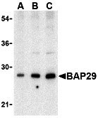
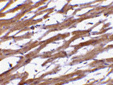
| Application Notes | Bap29 antibody can be used for the detection of Bap29 by Western blot at 0.5 - 2 µg/mL. Antibody can also be used for immunohistochemistry starting at 10 µg/mL. For immunofluorescence start at 20 µg/mL. |
| Gene ID | 55973 |
|---|---|
| Other Names | BAP29 Antibody: BAP29, BAP29, B-cell receptor-associated protein 29, BCR-associated protein 29, B-cell receptor-associated protein 29 |
| Target/Specificity | BCAP29; |
| Reconstitution & Storage | BAP29 antibody can be stored at 4℃ for three months and -20℃, stable for up to one year. As with all antibodies care should be taken to avoid repeated freeze thaw cycles. Antibodies should not be exposed to prolonged high temperatures. |
| Precautions | BAP29 Antibody is for research use only and not for use in diagnostic or therapeutic procedures. |
| Name | BCAP29 |
|---|---|
| Synonyms | BAP29 |
| Function | May play a role in anterograde transport of membrane proteins from the endoplasmic reticulum to the Golgi. May be involved in CASP8- mediated apoptosis (By similarity). |
| Cellular Location | Endoplasmic reticulum membrane; Multi-pass membrane protein |

Thousands of laboratories across the world have published research that depended on the performance of antibodies from Abcepta to advance their research. Check out links to articles that cite our products in major peer-reviewed journals, organized by research category.
info@abcepta.com, and receive a free "I Love Antibodies" mug.

Provided below are standard protocols that you may find useful for product applications.
Background
BAP29 Antibody: Bap29 and the related protein Bap31 are endoplasmic reticulum (ER) and ER-vesicle membrane proteins and members of the B-cell receptor-associated protein family. These two proteins are highly homologous and can form homo- and heterodimers. Both Bap29 and Bap31 interact with membrane-bound immunoglobulins (mIgs), such as IgM and IgD, which with Ig-alpha/Ig-beta heterodimers form B cell antigen receptors. Binding of the Bap29/Bap31 heterodimer correlates with the ER retention of non-Ig-alpha/Ig-beta bound mIg complexes, suggesting that Bap29 and Bap31 may act as chaperones transmembrane regions of various proteins. Bap29 possesses multiple isoforms.
References
Kim KM, Adachi T, Nielsen PJ, et al. Two new proteins preferentially associated with membrane immunoglobulin D. EMBO J.1994; 13:3793-800.
Ng F, Nguyen M, Kwan T, et al. p28 BAP31, a Bcl-2/Bcl-XL- and procaspase-8-associated protein the endoplasmic reticulum. J. Cell Biol.1997; 139:327-38.
Schamel WW, Kuppig S, Becker B, et al. A high-molecular-weight complex of membrane proteins BAP29/BAP31 is involved in the retention of membrane-bound IgD in the endoplasmic reticulum. Proc. Natl. Acad. Sci. USA2003; 100:9861-6.
If you have used an Abcepta product and would like to share how it has performed, please click on the "Submit Review" button and provide the requested information. Our staff will examine and post your review and contact you if needed.
If you have any additional inquiries please email technical services at tech@abcepta.com.
Ordering Information
Other Products
Shipping Information
