


 Foundational characteristics of cancer include proliferation, angiogenesis, migration, evasion of apoptosis, and cellular immortality. Find key markers for these cellular processes and antibodies to detect them.
 The SUMOplot™ Analysis Program predicts and scores sumoylation sites in your protein. SUMOylation is a post-translational modification involved in various cellular processes, such as nuclear-cytosolic transport, transcriptional regulation, apoptosis, protein stability, response to stress, and progression through the cell cycle.
 The Autophagy Receptor Motif Plotter predicts and scores autophagy receptor binding sites in your protein. Identifying proteins connected to this pathway is critical to understanding the role of autophagy in physiological as well as pathological processes such as development, differentiation, neurodegenerative diseases, stress, infection, and cancer.
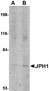
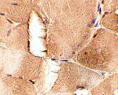
JPH1 Antibody
- SPECIFICATION
- CITATIONS
- PROTOCOLS
- BACKGROUND

Application
| WB, IHC-P, E |
|---|---|
| Primary Accession | Q9HDC5 |
| Other Accession | AAI39833, 145337941 |
| Reactivity | Human, Mouse, Rat |
| Host | Rabbit |
| Clonality | Polyclonal |
| Isotype | IgG |
| Calculated MW | 71686 Da |
| Application Notes | JPH1 antibody can be used for detection of JPH1 by Western blot at 1 - 2 µg/mL. Antibody can also be used for immunohistochemistry starting at 2.5 µg/mL. |
| Gene ID | 56704 |
|---|---|
| Target/Specificity | JPH1; |
| Reconstitution & Storage | JPH1 antibody can be stored at 4℃ for three months and -20℃, stable for up to one year. As with all antibodies care should be taken to avoid repeated freeze thaw cycles. Antibodies should not be exposed to prolonged high temperatures. |
| Precautions | JPH1 Antibody is for research use only and not for use in diagnostic or therapeutic procedures. |
| Name | JPH1 |
|---|---|
| Synonyms | JP1 |
| Function | Junctophilins contribute to the formation of junctional membrane complexes (JMCs) which link the plasma membrane with the endoplasmic or sarcoplasmic reticulum in excitable cells. Provides a structural foundation for functional cross-talk between the cell surface and intracellular calcium release channels. JPH1 contributes to the construction of the skeletal muscle triad by linking the t-tubule (transverse-tubule) and SR (sarcoplasmic reticulum) membranes. |
| Cellular Location | Cell membrane; Peripheral membrane protein. Endoplasmic reticulum membrane; Single-pass type IV membrane protein. Sarcoplasmic reticulum membrane; Single-pass type IV membrane protein. Note=Localized predominantly on the plasma membrane. The transmembrane domain is anchored in endoplasmic/sarcoplasmic reticulum membrane, while the N-terminal part associates with the plasma membrane. In skeletal muscle cells, it is predominantly localized at the junction of the A and I bands (By similarity). |
| Tissue Location | Abundantly expressed in skeletal muscle. Very low levels in heart. |

Thousands of laboratories across the world have published research that depended on the performance of antibodies from Abcepta to advance their research. Check out links to articles that cite our products in major peer-reviewed journals, organized by research category.
info@abcepta.com, and receive a free "I Love Antibodies" mug.

Provided below are standard protocols that you may find useful for product applications.
Background
JPH1 Antibody: Junctional complexes between the plasma membrane (PM) and endoplasmic/sarcoplasmic reticulum (ER/SR) are a common feature of all excitable cell types and mediate cross talk between cell surface and intracellular ion channels. Junctophilins (JPs) are important components of the junctional complexes. JPs are composed of a carboxy-terminal hydrophobic segment spanning the ER/SR membrane and a remaining cytoplasmic domain that shows specific affinity for the PM. Four JPs have been identified as tissue-specific subtypes derived from different genes: JPH1 is expressed in skeletal muscle, JPH2 is detected throughout all muscle cell types, and JPH3 and JPH4 are predominantly expressed in the brain and contribute to the subsurface cistern formation in neurons. JPH1 is essential for stabilizing the T-tubule and SR membranes to form junctions and provide an environment for the assembly of receptors such as the ryanodine receptor type 1 (RyR1).
References
Takeshima H, Komazaki S, Nishi M, et al. Junctophilins: a novel family of junctional membrane complex proteins. Mol. Cell.2000; 6:11-22.
Kakizawa S, Kishimoto Y, Hashimoto K, et al. Junctophilin-mediated channel crosstalk essential for cerebellar synaptic plasticity. EMBO J.2007; 26:1924-33.
Nishi M, Sakagami H, Komazaki S, et al. Coexpression of junctophilin type 3 and type 4 in brain. Brain Res. Mol. Brain Res.2003; 118:102-10.
Phimister AJ, Lango J, Lee EH, et al. Conformation-dependent stability of Junctophilin 1 (JP1) and Ryanodine Receptor type 1 (RyR1) channel complex is mediated by their hyper-reactive thiols. J. Biol. Chem.2007; 282:8867-77.
If you have used an Abcepta product and would like to share how it has performed, please click on the "Submit Review" button and provide the requested information. Our staff will examine and post your review and contact you if needed.
If you have any additional inquiries please email technical services at tech@abcepta.com.
Ordering Information
Other Products
Shipping Information
