


 Foundational characteristics of cancer include proliferation, angiogenesis, migration, evasion of apoptosis, and cellular immortality. Find key markers for these cellular processes and antibodies to detect them.
 The SUMOplot™ Analysis Program predicts and scores sumoylation sites in your protein. SUMOylation is a post-translational modification involved in various cellular processes, such as nuclear-cytosolic transport, transcriptional regulation, apoptosis, protein stability, response to stress, and progression through the cell cycle.
 The Autophagy Receptor Motif Plotter predicts and scores autophagy receptor binding sites in your protein. Identifying proteins connected to this pathway is critical to understanding the role of autophagy in physiological as well as pathological processes such as development, differentiation, neurodegenerative diseases, stress, infection, and cancer.
JMJD2A Antibody
- SPECIFICATION
- CITATIONS
- PROTOCOLS
- BACKGROUND

Application
| WB, IHC-P, E |
|---|---|
| Primary Accession | O75164 |
| Other Accession | CAH71021, 55665236 |
| Reactivity | Human, Mouse, Rat |
| Host | Rabbit |
| Clonality | Polyclonal |
| Isotype | IgG |
| Calculated MW | 120662 Da |
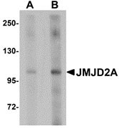
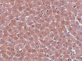
| Application Notes | JMJD2A antibody can be used for detection of JMJD2A by Western blot at 1 - 2 µg/mL. Antibody can also be used for immunohistochemistry starting at 5 µg/mL. |
| Gene ID | 9682 |
|---|---|
| Target/Specificity | KDM4A; |
| Reconstitution & Storage | JMJD2A antibody can be stored at 4℃ for three months and -20℃, stable for up to one year. As with all antibodies care should be taken to avoid repeated freeze thaw cycles. Antibodies should not be exposed to prolonged high temperatures. |
| Precautions | JMJD2A Antibody is for research use only and not for use in diagnostic or therapeutic procedures. |
| Name | KDM4A |
|---|---|
| Synonyms | JHDM3A, JMJD2, JMJD2A, KIAA0677 |
| Function | Histone demethylase that specifically demethylates 'Lys-9' and 'Lys-36' residues of histone H3, thereby playing a central role in histone code (PubMed:26741168, PubMed:21768309). Does not demethylate histone H3 'Lys-4', H3 'Lys-27' nor H4 'Lys-20'. Demethylates trimethylated H3 'Lys-9' and H3 'Lys-36' residue, while it has no activity on mono- and dimethylated residues. Demethylation of Lys residue generates formaldehyde and succinate. Participates in transcriptional repression of ASCL2 and E2F-responsive promoters via the recruitment of histone deacetylases and NCOR1, respectively. |
| Cellular Location | Nucleus {ECO:0000255|PROSITE-ProRule:PRU00537, ECO:0000269|PubMed:15927959, ECO:0000269|PubMed:16024779} |
| Tissue Location | Ubiquitous.. |

Thousands of laboratories across the world have published research that depended on the performance of antibodies from Abcepta to advance their research. Check out links to articles that cite our products in major peer-reviewed journals, organized by research category.
info@abcepta.com, and receive a free "I Love Antibodies" mug.

Provided below are standard protocols that you may find useful for product applications.
Background
JMJD2A Antibody: Members of the Jumonji domain 2 (JMJD2) family contain a JmjN domain, a JmjC domain, a JD2H domain, two TUDOR domains, and two PHD-type zinc fingers. The first member of this group, JMJD2A, is widely expressed in human tissues and cell lines and functions as a trimethylation-specific demethylase, converting specific trimethylated histone residues to the dimethylated form, and as a transcriptional repressor. JMJD2A can also form a complex with the androgen receptor (AR), a transcription factor that is pivotal for the development of prostate cancer. Overexpression of JMJD2A stimulates AR function and this stimulation is dependent on JMJD2A catalytic activity, suggesting that JMJD2A might be a critical protein with roles in cell proliferation and oncogenesis.
References
Gray SG, Iglesias AH, Lizcano F, et al. Functional characterization of JMJD2A, a histone deacetylase- and retinoblastoma-binding protein. J. Biol. Chem.2005; 31:28507-18.
Shin S and Janknecht R. Activation of androgen receptor by histone demethylases JMJD2A and JMJD2D. Biochem. Biophys. Res. Comm.2007; 359:742-6.
If you have used an Abcepta product and would like to share how it has performed, please click on the "Submit Review" button and provide the requested information. Our staff will examine and post your review and contact you if needed.
If you have any additional inquiries please email technical services at tech@abcepta.com.
Ordering Information
Other Products
Shipping Information
