


 Foundational characteristics of cancer include proliferation, angiogenesis, migration, evasion of apoptosis, and cellular immortality. Find key markers for these cellular processes and antibodies to detect them.
 The SUMOplot™ Analysis Program predicts and scores sumoylation sites in your protein. SUMOylation is a post-translational modification involved in various cellular processes, such as nuclear-cytosolic transport, transcriptional regulation, apoptosis, protein stability, response to stress, and progression through the cell cycle.
 The Autophagy Receptor Motif Plotter predicts and scores autophagy receptor binding sites in your protein. Identifying proteins connected to this pathway is critical to understanding the role of autophagy in physiological as well as pathological processes such as development, differentiation, neurodegenerative diseases, stress, infection, and cancer.
EFHD1 Antibody
- SPECIFICATION
- CITATIONS
- PROTOCOLS
- BACKGROUND

Application
| WB, IF, E |
|---|---|
| Primary Accession | Q9BUP0 |
| Other Accession | NP_079478, 20149496 |
| Reactivity | Human, Mouse, Rat |
| Host | Rabbit |
| Clonality | Polyclonal |
| Isotype | IgG |
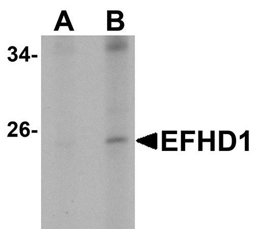
| Calculated MW | 26928 Da |
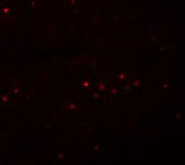
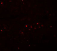
| Application Notes | EFHD1 antibody can be used for detection of EFHD1 by Western blot at 2 - 4 µg/mL. Antibody can also be used for immunoflourescence starting at 5 µg/mL. For immunofluorescence start at 20 µg/mL. |
| Gene ID | 80303 |
|---|---|
| Target/Specificity | EFHD1; |
| Reconstitution & Storage | EFHD1 antibody can be stored at 4℃ for three months and -20℃, stable for up to one year. As with all antibodies care should be taken to avoid repeated freeze thaw cycles. Antibodies should not be exposed to prolonged high temperatures. |
| Precautions | EFHD1 Antibody is for research use only and not for use in diagnostic or therapeutic procedures. |
| Name | EFHD1 |
|---|---|
| Synonyms | SWS2 |
| Function | Acts as a calcium sensor for mitochondrial flash (mitoflash) activation, an event characterized by stochastic bursts of superoxide production (PubMed:26975899). May play a role in neuronal differentiation (By similarity). |
| Cellular Location | Mitochondrion inner membrane {ECO:0000250|UniProtKB:Q9D4J1} |

Thousands of laboratories across the world have published research that depended on the performance of antibodies from Abcepta to advance their research. Check out links to articles that cite our products in major peer-reviewed journals, organized by research category.
info@abcepta.com, and receive a free "I Love Antibodies" mug.

Provided below are standard protocols that you may find useful for product applications.
Background
EFHD1 Antibody: EFHD1, also known as Swiprosin-2 or SWS2, is an EF-hand and coiled-coil-containing adaptor protein identified in a subtractive hybridization study using a neuronal cell line established from the cerebellum of an adult p53-null mouse, however further study indicated no difference in normal mice. Its mRNA is widely expressed, with its expression in brain undetectable at embryonic stages, with increasing levels from postnatal to adult development. In situ hybridization showed expression in neurons but not white matter of the cerebellum and cerebrum. EHFD1 is also highly expressed in testes, ovary, and the collecting ducts of the kidney, suggesting that in non-neuronal cells, EFHD1 may be involved in gametogenesis and water-reabsorbtion.
References
Tominaga M and Tomooka Y. Novel genes cloned from a neuronal cell line newly established from a cerebellum of an adult p53-/- mouse. Biochem. Biophys. Res. Comm.2002; 297:473-9.
If you have used an Abcepta product and would like to share how it has performed, please click on the "Submit Review" button and provide the requested information. Our staff will examine and post your review and contact you if needed.
If you have any additional inquiries please email technical services at tech@abcepta.com.
Ordering Information
Other Products
Shipping Information
