


 Foundational characteristics of cancer include proliferation, angiogenesis, migration, evasion of apoptosis, and cellular immortality. Find key markers for these cellular processes and antibodies to detect them.
 The SUMOplot™ Analysis Program predicts and scores sumoylation sites in your protein. SUMOylation is a post-translational modification involved in various cellular processes, such as nuclear-cytosolic transport, transcriptional regulation, apoptosis, protein stability, response to stress, and progression through the cell cycle.
 The Autophagy Receptor Motif Plotter predicts and scores autophagy receptor binding sites in your protein. Identifying proteins connected to this pathway is critical to understanding the role of autophagy in physiological as well as pathological processes such as development, differentiation, neurodegenerative diseases, stress, infection, and cancer.
FAM82A1 Antibody
- SPECIFICATION
- CITATIONS
- PROTOCOLS
- BACKGROUND

Application
| WB, IF, ICC, E |
|---|---|
| Primary Accession | Q96LZ7 |
| Other Accession | NP_653314, 283046686 |
| Reactivity | Human, Mouse, Rat |
| Host | Chicken |
| Clonality | Polyclonal |
| Isotype | IgY |
| Calculated MW | 47399 Da |
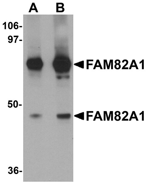
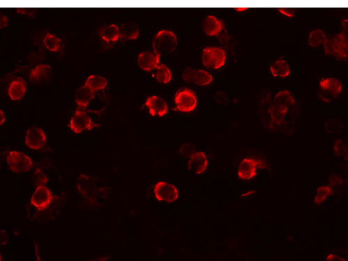
| Application Notes | FAM82A1 antibody can be used for detection of FAM82A1 by Western blot at 1 - 2 µg/mL. Antibody can also be used for immunocytochemistry starting at 5 µg/mL. For immunofluorescence start at 20 µg/mL. |
| Gene ID | 151393 |
|---|---|
| Target/Specificity | FAM82A1; |
| Reconstitution & Storage | FAM82A1 antibody can be stored at 4℃ for three months and -20℃, stable for up to one year. As with all antibodies care should be taken to avoid repeated freeze thaw cycles. Antibodies should not be exposed to prolonged high temperatures. |
| Precautions | FAM82A1 Antibody is for research use only and not for use in diagnostic or therapeutic procedures. |
| Name | RMDN2 |
|---|---|
| Synonyms | FAM82A, FAM82A1 |
| Cellular Location | Membrane; Single-pass membrane protein. Cytoplasm Cytoplasm, cytoskeleton, spindle Cytoplasm, cytoskeleton, spindle pole Note=In interphase localizes in the cytoplasm, and during mitosis localizes to the spindle microtubules and spindle poles. Also detected as large dots in the perinuclear region |

Thousands of laboratories across the world have published research that depended on the performance of antibodies from Abcepta to advance their research. Check out links to articles that cite our products in major peer-reviewed journals, organized by research category.
info@abcepta.com, and receive a free "I Love Antibodies" mug.

Provided below are standard protocols that you may find useful for product applications.
Background
FAM82A1 Antibody: The human protein FAM82A1, also known as regulator of microtubule dynamics 2 (RMD2), was one of three proteins identified as related to a family of microtubule-associated proteins in C. elegans. FAM82A1 contains multiple coiled-coil domains and localize to the spindle microtubules and spindle poles during cell division. During interphase, RMD2 localizes to the cytoplasm with protein observed in the microtubule lattice and perinuclear region.
References
Oishi K, Okanu H, and Sawa H. RMD-1, a novel microtubule-associated protein, functions in chromosome segregation in Caenorhabditis elegans. J. Cell Biol.2007; 179:1149-62.
If you have used an Abcepta product and would like to share how it has performed, please click on the "Submit Review" button and provide the requested information. Our staff will examine and post your review and contact you if needed.
If you have any additional inquiries please email technical services at tech@abcepta.com.
Ordering Information
Other Products
Shipping Information
