


 Foundational characteristics of cancer include proliferation, angiogenesis, migration, evasion of apoptosis, and cellular immortality. Find key markers for these cellular processes and antibodies to detect them.
 The SUMOplot™ Analysis Program predicts and scores sumoylation sites in your protein. SUMOylation is a post-translational modification involved in various cellular processes, such as nuclear-cytosolic transport, transcriptional regulation, apoptosis, protein stability, response to stress, and progression through the cell cycle.
 The Autophagy Receptor Motif Plotter predicts and scores autophagy receptor binding sites in your protein. Identifying proteins connected to this pathway is critical to understanding the role of autophagy in physiological as well as pathological processes such as development, differentiation, neurodegenerative diseases, stress, infection, and cancer.
APBA2 Antibody
- SPECIFICATION
- CITATIONS
- PROTOCOLS
- BACKGROUND

Application
| WB, IHC-P, IF, E |
|---|---|
| Primary Accession | Q99767 |
| Other Accession | NP_005494, 22035550 |
| Reactivity | Human |
| Host | Rabbit |
| Clonality | Polyclonal |
| Isotype | IgG |
| Calculated MW | 82512 Da |
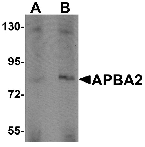
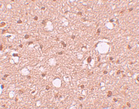
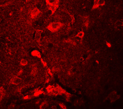
| Application Notes | APBA2 antibody can be used for detection of APBA2 by Western blot at 1 - 2 µg/mL. Antibody can also be used for immunohistochemistry starting at 10 µg/mL. For immunofluorescence start at 20 µg/mL. |
| Gene ID | 321 |
|---|---|
| Target/Specificity | APBA2; |
| Reconstitution & Storage | APBA2 antibody can be stored at 4℃ for three months and -20℃, stable for up to one year. As with all antibodies care should be taken to avoid repeated freeze thaw cycles. Antibodies should not be exposed to prolonged high temperatures. |
| Precautions | APBA2 Antibody is for research use only and not for use in diagnostic or therapeutic procedures. |
| Name | APBA2 |
|---|---|
| Synonyms | MINT2, X11L |
| Function | Putative function in synaptic vesicle exocytosis by binding to STXBP1, an essential component of the synaptic vesicle exocytotic machinery. May modulate processing of the amyloid-beta precursor protein (APP) and hence formation of APP-beta. |
| Tissue Location | Brain. |

Thousands of laboratories across the world have published research that depended on the performance of antibodies from Abcepta to advance their research. Check out links to articles that cite our products in major peer-reviewed journals, organized by research category.
info@abcepta.com, and receive a free "I Love Antibodies" mug.

Provided below are standard protocols that you may find useful for product applications.
Background
APBA2 Antibody: APBA2, a member of the X11 protein family, is a phosphotyrosine-binding domain protein and is a neuronal adapter protein that interacts with amyloid precursor protein (APP) and neuritic plaques in the brains of patients with Alzheimer's disease. It stabilizes APP and inhibits production of proteolytic APP fragments including the Abeta peptide that is deposited in the brains of Alzheimer's disease patients. APBA2 is believed to be involved in signal transduction processes and is also regarded as a putative vesicular trafficking protein in the brain that can form a complex with the potential to couple synaptic vesicle exocytosis to neuronal cell adhesion. Recent reports suggest that it may also be a candidate gene for autism.
References
Blanco G, Irving NG, Brown SDM, et al. Mapping of the human and murine X11-like genes (APBA2 and Apba2), the murine Fe65 gene (Apbb1), and the human Fe65-like gene (APBB2): genes encoding phosphotyrosine-binding domain proteins that interact with the Alzheimer’s disease amyloid precursor protein. Mamm. Gen.1988; 9:473-5.
Mint2/X11-like colocalizes with the Alzheimer’s disease amyloid precursor protein and is associated with neuritic plaques in Alzheimer’s disease. Eur. J. Neurosci.1999; 11:1988-94.
Lee JH, Lau KF, Perkinton MS, et al. The neuronal adaptor protein X11beta reduces amyloid beta-protein levels and amyloid plaque formation in the brains of transgenic mice. J. Biol. Chem.2004; 279:49099-104.
Lee HW, Park JW, Sandagsuren EU, et al. Overexpression of APP stimulates basal and constitutive exocytosis in PC12 cells. Neurosci. Lett.2008; 436:245-9.
If you have used an Abcepta product and would like to share how it has performed, please click on the "Submit Review" button and provide the requested information. Our staff will examine and post your review and contact you if needed.
If you have any additional inquiries please email technical services at tech@abcepta.com.
Ordering Information
Other Products
Shipping Information
