


 Foundational characteristics of cancer include proliferation, angiogenesis, migration, evasion of apoptosis, and cellular immortality. Find key markers for these cellular processes and antibodies to detect them.
 The SUMOplot™ Analysis Program predicts and scores sumoylation sites in your protein. SUMOylation is a post-translational modification involved in various cellular processes, such as nuclear-cytosolic transport, transcriptional regulation, apoptosis, protein stability, response to stress, and progression through the cell cycle.
 The Autophagy Receptor Motif Plotter predicts and scores autophagy receptor binding sites in your protein. Identifying proteins connected to this pathway is critical to understanding the role of autophagy in physiological as well as pathological processes such as development, differentiation, neurodegenerative diseases, stress, infection, and cancer.
IRF2BP2 Antibody
- SPECIFICATION
- CITATIONS
- PROTOCOLS
- BACKGROUND

Application
| WB, ICC, E |
|---|---|
| Primary Accession | Q7Z5L9 |
| Other Accession | NP_892017, 116734704 |
| Reactivity | Human, Mouse |
| Host | Rabbit |
| Clonality | Polyclonal |
| Isotype | IgG |
| Calculated MW | 61025 Da |
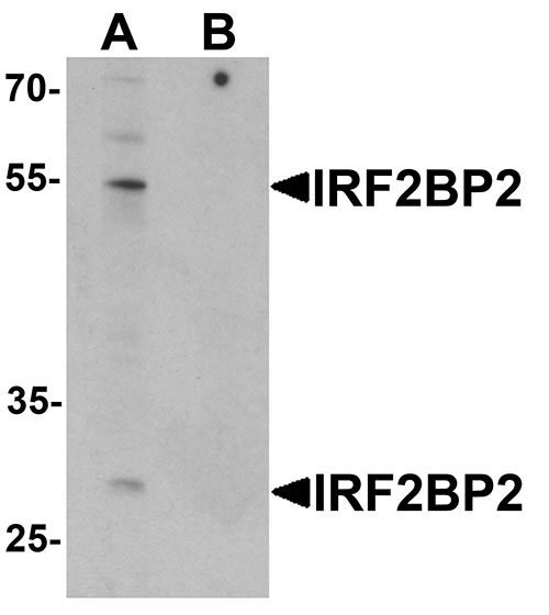
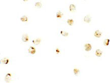
| Application Notes | IRF2BP2 antibody can be used for detection of IRF2BP2 by Western blot at 1 µg/mL. Antibody can also be used for immunocytochemistry starting at 2.5 µg/mL. |
| Gene ID | 359948 |
|---|---|
| Target/Specificity | IRF2BP2; At least two isoforms of IRF2BP2 are known to exist; this antibody will detect both. This antibody is predicted to not cross-react with any FOXD4 protein family members. |
| Reconstitution & Storage | IRF2BP2 antibody can be stored at 4℃ for three months and -20℃, stable for up to one year. As with all antibodies care should be taken to avoid repeated freeze thaw cycles. Antibodies should not be exposed to prolonged high temperatures. |
| Precautions | IRF2BP2 Antibody is for research use only and not for use in diagnostic or therapeutic procedures. |
| Name | IRF2BP2 |
|---|---|
| Function | Acts as a transcriptional corepressor in a IRF2-dependent manner; this repression is not mediated by histone deacetylase activities (PubMed:12799427). Represses the NFAT1-dependent transactivation of NFAT-responsive promoters (PubMed:21576369). Acts as a coactivator of VEGFA expression in cardiac and skeletal muscles (PubMed:20702774). Plays a role in immature B-cell differentiation (PubMed:27016798). |
| Cellular Location | Cytoplasm. Nucleus. |

Thousands of laboratories across the world have published research that depended on the performance of antibodies from Abcepta to advance their research. Check out links to articles that cite our products in major peer-reviewed journals, organized by research category.
info@abcepta.com, and receive a free "I Love Antibodies" mug.

Provided below are standard protocols that you may find useful for product applications.
Background
IRF2BP2 Antibody: IRF2BP2, like the related protein IRF2BP1, is a co-repressor that interacts specifically with the C-terminal repression domain of Interferon Regulatory Factor 2 (IRF2). IRF2BP2 is a direct target gene of p53 and is involved in cell survival during the p53 stress response, able to impede the p53-mediated transactivation of p21 and Bax. IRF2BP2 is also a co-factor of VGLL4 and is required to induce the expression of vascular endothelial growth factor A (VEGF-A) in muscle. It is normally found in the nucleus of skeletal muscle and cardiac cells, but can be found in the cytoplasm during skeletal muscle differentiation.
References
Childs KS and Goodbourn S. Identification of novel co-repressor molecules for interferon regulatory factor-2. Nuc. Acids Res. 2003; 31:3016-26
Koeppel M, van Heeringen SJ, Smeenk L, et al. The novel p53 target gene IRF2BP2 participates in cell survival during the p53 stress response. Nuc. Acids Res. 2009; 37:322-35.
Teng ACT, Kuraitis D, Deeke SA, et al. IRF2BP2 is a skeletal and cardiac muscle-enriched ischemia-inducible activator of VEGFA expression. FASEB J. 2010; 24:4825-34.
Teng ACT, Al-montashiri NAM, Cheng BLM, et al. Identification of a phosphorylation-dependent nuclear localization motif in interferon regulatory factor 2 binding protein 2. PLoS One 2011; 6:e24100.
If you have used an Abcepta product and would like to share how it has performed, please click on the "Submit Review" button and provide the requested information. Our staff will examine and post your review and contact you if needed.
If you have any additional inquiries please email technical services at tech@abcepta.com.
Ordering Information
Other Products
Shipping Information
