


 Foundational characteristics of cancer include proliferation, angiogenesis, migration, evasion of apoptosis, and cellular immortality. Find key markers for these cellular processes and antibodies to detect them.
 The SUMOplot™ Analysis Program predicts and scores sumoylation sites in your protein. SUMOylation is a post-translational modification involved in various cellular processes, such as nuclear-cytosolic transport, transcriptional regulation, apoptosis, protein stability, response to stress, and progression through the cell cycle.
 The Autophagy Receptor Motif Plotter predicts and scores autophagy receptor binding sites in your protein. Identifying proteins connected to this pathway is critical to understanding the role of autophagy in physiological as well as pathological processes such as development, differentiation, neurodegenerative diseases, stress, infection, and cancer.
RNASET2 Antibody
- SPECIFICATION
- CITATIONS
- PROTOCOLS
- BACKGROUND

Application
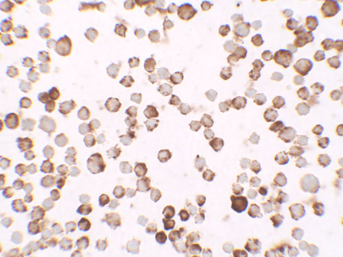
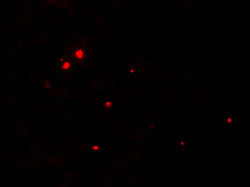
| WB, IF, ICC, E |
|---|---|
| Primary Accession | O00584 |
| Other Accession | NP_003721, 5231228 |
| Reactivity | Human, Mouse, Rat |
| Host | Rabbit |
| Clonality | Polyclonal |
| Isotype | IgG |
| Calculated MW | 29481 Da |
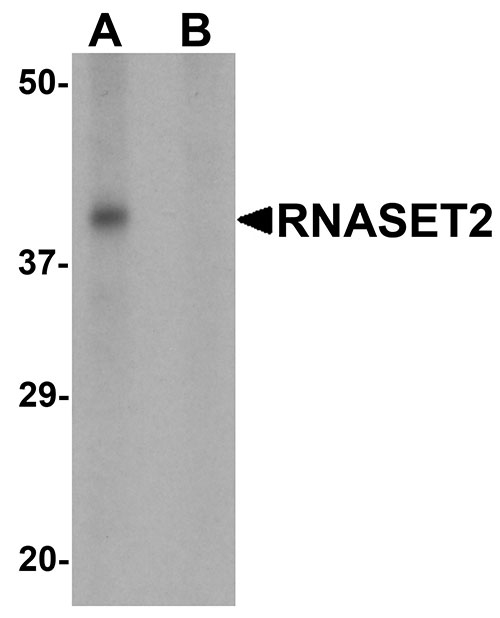
| Application Notes | RNASET2 antibody can be used for detection of FOXRED2 by Western blot at 1 µg/mL. |
| Gene ID | 8635 |
|---|---|
| Target/Specificity | RNASET2; |
| Reconstitution & Storage | RNASET2 antibody can be stored at 4℃ for three months and -20℃, stable for up to one year. As with all antibodies care should be taken to avoid repeated freeze thaw cycles. Antibodies should not be exposed to prolonged high temperatures. |
| Precautions | RNASET2 Antibody is for research use only and not for use in diagnostic or therapeutic procedures. |
| Name | RNASET2 |
|---|---|
| Synonyms | RNASE6PL |
| Function | Ribonuclease that plays an essential role in innate immune response by recognizing and degrading RNAs from microbial pathogens that are subsequently sensed by TLR8 (PubMed:31778653). Cleaves preferentially single-stranded RNA molecules between purine and uridine residues, which critically contributes to the supply of catabolic uridine and the generation of purine-2',3'-cyclophosphate-terminated oligoribonucleotides (PubMed:31778653, PubMed:38697119). In turn, RNase T2 degradation products promote the RNA-dependent activation of TLR8 (PubMed:31778653). In plasmacytoid dendritic cells, it cooperates with PLD3 or PLD4 5'->3' exonucleases to process RNA fragments and release 2',3'-cyclic guanosine monophosphate (2',3'-cGMP), a potent stimulatory ligand for TLR7 (PubMed:38697119). Also plays a key role in degradation of mitochondrial RNA and processing of non-coding RNA imported from the cytosol into mitochondria (PubMed:28730546, PubMed:30184494). Participates as well in degradation of mitochondrion-associated cytosolic rRNAs (PubMed:30385512). |
| Cellular Location | Secreted. Lysosome lumen. Endoplasmic reticulum lumen. Mitochondrion intermembrane space. Note=Full-length RNASET2 is found in the endoplasmic reticulum while smaller RNASET2 proteolytic products are found in the lysosome fraction. |
| Tissue Location | Ubiquitous. Higher expression levels observed in the temporal lobe and fetal brain. |

Thousands of laboratories across the world have published research that depended on the performance of antibodies from Abcepta to advance their research. Check out links to articles that cite our products in major peer-reviewed journals, organized by research category.
info@abcepta.com, and receive a free "I Love Antibodies" mug.

Provided below are standard protocols that you may find useful for product applications.
Background
RNASET2 Antibody: RNASET2 is a novel member of the Rh/T2/S-glycoprotein class of extracellular ribonucleases. It is a single copy gene that maps to 6q27, a region associated with human malignancies and chromosomal rearrangement, and has been suggested to function as a tumor suppressor protein. Its expression is suppressed in Human T-cell Leukemia Virus type 1 (HTLV-1) infected cells following the binding of the HTLV-1 Tax protein to the RNASET2 promoter. As Adult T-cell leukemia (ATL) is one of the primary diseases caused by HTLV-1 infection, a reduction in the level of RNASET2 by Tax may play a role in ATL development.
References
Acquati F, Morelli C, Cinquetti R, et al. Cloning and characterization of a senescence inducing and class II tumor suppressor gene in ovarian carcinoma at chromosome region 6q27. Oncogene 2001; 20:980-8.
Campomenosi P, Salis S, Lingqvist C, et al. Characterization of RNASET2, the first human member of the Rh/T2/S family of glycoproteins. Arch. Biochm. Biophys. 2006; 449:17-26
Polakowski N, Han H, and Lemasson I. Direct inhibition of RNase T2 expression by the HLTV-1 viral protein Tax. Viruses 2011; 3:1485-500.
If you have used an Abcepta product and would like to share how it has performed, please click on the "Submit Review" button and provide the requested information. Our staff will examine and post your review and contact you if needed.
If you have any additional inquiries please email technical services at tech@abcepta.com.
Ordering Information
Other Products
Shipping Information
