


 Foundational characteristics of cancer include proliferation, angiogenesis, migration, evasion of apoptosis, and cellular immortality. Find key markers for these cellular processes and antibodies to detect them.
 The SUMOplot™ Analysis Program predicts and scores sumoylation sites in your protein. SUMOylation is a post-translational modification involved in various cellular processes, such as nuclear-cytosolic transport, transcriptional regulation, apoptosis, protein stability, response to stress, and progression through the cell cycle.
 The Autophagy Receptor Motif Plotter predicts and scores autophagy receptor binding sites in your protein. Identifying proteins connected to this pathway is critical to understanding the role of autophagy in physiological as well as pathological processes such as development, differentiation, neurodegenerative diseases, stress, infection, and cancer.
YPEL5 Antibody
- SPECIFICATION
- CITATIONS
- PROTOCOLS
- BACKGROUND

Application
| WB, IF, ICC, E |
|---|---|
| Primary Accession | P62699 |
| Other Accession | NP_057145, 7706341 |
| Reactivity | Human, Mouse |
| Host | Rabbit |
| Clonality | Polyclonal |
| Isotype | IgG |
| Calculated MW | 13842 Da |
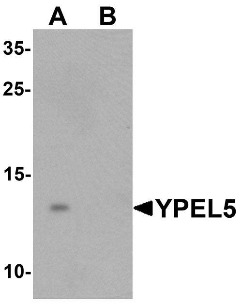
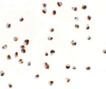
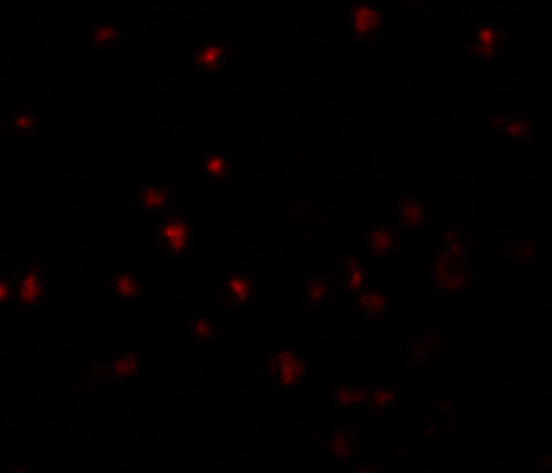
| Application Notes | YPEL5 antibody can be used for detection of YPEL5 by Western blot at 1 µg/mL. Antibody can also be used for immunocytochemistry starting at 2.5 µg/mL. For immunofluorescence start at 2.5 µg/mL. |
| Gene ID | 51646 |
|---|---|
| Target/Specificity | YPEL5; |
| Reconstitution & Storage | YPEL5 antibody can be stored at 4℃ for three months and -20℃, stable for up to one year. As with all antibodies care should be taken to avoid repeated freeze thaw cycles. Antibodies should not be exposed to prolonged high temperatures. |
| Precautions | YPEL5 Antibody is for research use only and not for use in diagnostic or therapeutic procedures. |
| Name | YPEL5 |
|---|---|
| Function | Component of the CTLH E3 ubiquitin-protein ligase complex that selectively accepts ubiquitin from UBE2H and mediates ubiquitination and subsequent proteasomal degradation of the transcription factor HBP1 (PubMed:29911972). Required for normal cell proliferation (By similarity). |
| Cellular Location | Nucleus {ECO:0000250|UniProtKB:Q65Z55}. Cytoplasm, cytoskeleton, microtubule organizing center, centrosome {ECO:0000250|UniProtKB:Q65Z55}. Cytoplasm, cytoskeleton, spindle pole {ECO:0000250|UniProtKB:Q65Z55}. Midbody {ECO:0000250|UniProtKB:Q65Z55} Note=Deteted in nucleus and at the centrosome during interphase. During mitosis, detected on the mitotic spindle, at spindle poles and at the midbody. {ECO:0000250|UniProtKB:Q65Z55} |

Thousands of laboratories across the world have published research that depended on the performance of antibodies from Abcepta to advance their research. Check out links to articles that cite our products in major peer-reviewed journals, organized by research category.
info@abcepta.com, and receive a free "I Love Antibodies" mug.

Provided below are standard protocols that you may find useful for product applications.
Background
YPEL5 Antibody: YPEL5 (yippee-like 5) belongs to a family of five yippee-like proteins, all of which localize to the centrosome or mitotic spindle and are widely expressed in both adult and fetal tissue. This localization plus the fact that the family of human YPEL proteins share a high degree of sequence homology across species suggests that these proteins may have a conserved function involved in cell division. YPEL5 is expressed at the nucleus and centrosome during interphase; during mitosis, it localizes to the spindle poles, mitotic spindle, and spindle midzone during mitosis. Finally, during cytokinesis, YPEL5 is localized to the midbody. It is associated with the Ran Binding Protein in the Microtubule organizing center (RanBPM) and a related protein RanBP10.
References
Hosono K, Sasaki T, Minoshima S, et al. Identification and characterization of a novel gene family YPEL in a wide spectrum of eukaryotic species. Gene 2004; 340: 31-43.
Hosono K, Noda S, Shimuzu A, et al. YPEL5 protein of the YPEL gene family is involved in the cell cycle progression by interacting with two distinct proteins RanBPM and RanBP10. Genomics 2010; 96:102-11.
If you have used an Abcepta product and would like to share how it has performed, please click on the "Submit Review" button and provide the requested information. Our staff will examine and post your review and contact you if needed.
If you have any additional inquiries please email technical services at tech@abcepta.com.
Ordering Information
Other Products
Shipping Information
