


 Foundational characteristics of cancer include proliferation, angiogenesis, migration, evasion of apoptosis, and cellular immortality. Find key markers for these cellular processes and antibodies to detect them.
 The SUMOplot™ Analysis Program predicts and scores sumoylation sites in your protein. SUMOylation is a post-translational modification involved in various cellular processes, such as nuclear-cytosolic transport, transcriptional regulation, apoptosis, protein stability, response to stress, and progression through the cell cycle.
 The Autophagy Receptor Motif Plotter predicts and scores autophagy receptor binding sites in your protein. Identifying proteins connected to this pathway is critical to understanding the role of autophagy in physiological as well as pathological processes such as development, differentiation, neurodegenerative diseases, stress, infection, and cancer.
SEPT1 Antibody
- SPECIFICATION
- CITATIONS
- PROTOCOLS
- BACKGROUND

Application
| WB, IF, ICC, E |
|---|---|
| Primary Accession | Q8WYJ6 |
| Other Accession | NP_443070, 431822382 |
| Reactivity | Human, Mouse, Rat |
| Host | Rabbit |
| Clonality | Polyclonal |
| Isotype | IgG |
| Calculated MW | 42386 Da |
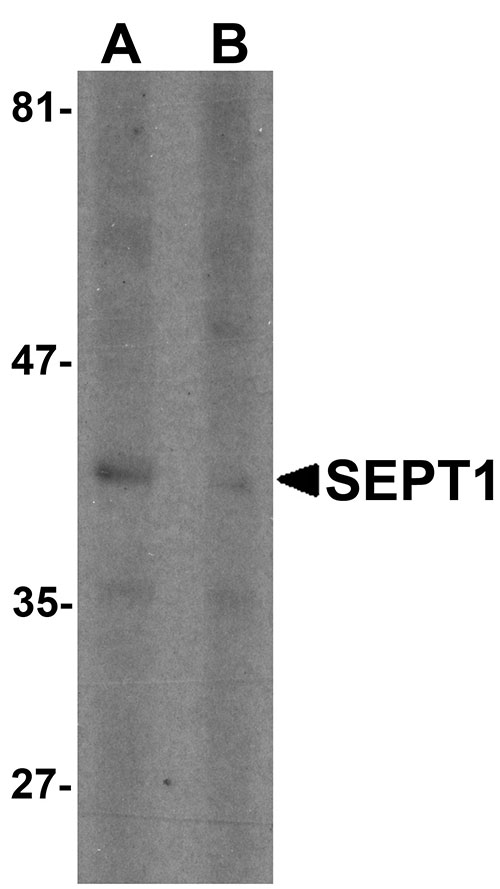
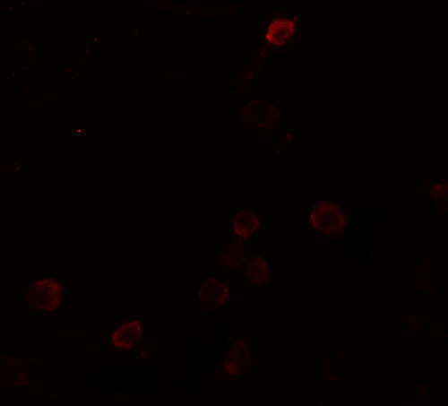
| Application Notes | SEPT1 antibody can be used for detection of SEPT1 by Western blot at 1 µg/mL. Antibody can also be used for immunocytochemistry starting at 5 µg/mL. For immunofluorescence start at 5 µg/mL. |
| Gene ID | 1731 |
|---|---|
| Target/Specificity | SEPT1; At least two isoforms of SEPT1 are known to exist. This antibody is predicted to not cross-react with other Septin protein family members. |
| Reconstitution & Storage | SEPT1 antibody can be stored at 4℃ for three months and -20℃, stable for up to one year. As with all antibodies care should be taken to avoid repeated freeze thaw cycles. Antibodies should not be exposed to prolonged high temperatures. |
| Precautions | SEPT1 Antibody is for research use only and not for use in diagnostic or therapeutic procedures. |
| Name | SEPTIN1 (HGNC:2879) |
|---|---|
| Synonyms | DIFF6, PNUTL3, SEPT1 |
| Function | Filament-forming cytoskeletal GTPase (By similarity). May play a role in cytokinesis (Potential). |
| Cellular Location | Cytoplasm. Cytoplasm, cytoskeleton. Cytoplasm, cytoskeleton, microtubule organizing center, centrosome. Midbody. Note=Remains at the centrosomes and the nearby microtubules throughout mitosis. Localizes to the midbody during cytokinesis |
| Tissue Location | Expressed at high levels in lymphoid and hematopoietic tissues. |

Thousands of laboratories across the world have published research that depended on the performance of antibodies from Abcepta to advance their research. Check out links to articles that cite our products in major peer-reviewed journals, organized by research category.
info@abcepta.com, and receive a free "I Love Antibodies" mug.

Provided below are standard protocols that you may find useful for product applications.
Background
SEPT1 Antibody: SEPT1 (also known as Septin 1), is a member of the septin family of GTPases. Members of this family are required for cytokinesis and the maintenance of cellular morphology. SEPT1 was initially identified through yeast two-hybrid screening as an interaction partner for the serine/threonine kinase Aurora-B. During mitosis, SEPT1 localizes to the spindle pole, suggesting that it may play a role in cytokinesis and chromosome congression.
References
Qi M, Yu W, Liu S, et al. Septin1, a new interaction partner for human serine/threonine kinase aurora-B. Biochem. Biophys. Res. Commun. 2005; 336:994-1000.
Russell SE and Hall PA. Septin genomics: a road less travelled. Biol. Chem. 2011; 392:763-7.
Zhu J, Qi ST, Wang YP, et al. Septin1 is required for spindle assembly and chromosome congression in mouse oocytes. Dev. Dyn. 2011; 240:2281-9.
If you have used an Abcepta product and would like to share how it has performed, please click on the "Submit Review" button and provide the requested information. Our staff will examine and post your review and contact you if needed.
If you have any additional inquiries please email technical services at tech@abcepta.com.
Ordering Information
Other Products
Shipping Information
