


 Foundational characteristics of cancer include proliferation, angiogenesis, migration, evasion of apoptosis, and cellular immortality. Find key markers for these cellular processes and antibodies to detect them.
 The SUMOplot™ Analysis Program predicts and scores sumoylation sites in your protein. SUMOylation is a post-translational modification involved in various cellular processes, such as nuclear-cytosolic transport, transcriptional regulation, apoptosis, protein stability, response to stress, and progression through the cell cycle.
 The Autophagy Receptor Motif Plotter predicts and scores autophagy receptor binding sites in your protein. Identifying proteins connected to this pathway is critical to understanding the role of autophagy in physiological as well as pathological processes such as development, differentiation, neurodegenerative diseases, stress, infection, and cancer.
TDGF1 Antibody
- SPECIFICATION
- CITATIONS
- PROTOCOLS
- BACKGROUND

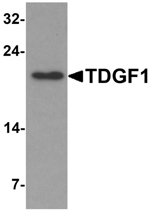
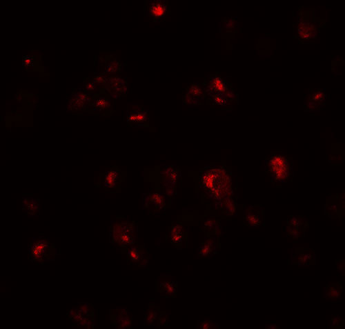
Application
| WB, IF, E |
|---|---|
| Primary Accession | P13385 |
| Other Accession | NP_003203, 4507425 |
| Reactivity | Human, Mouse, Rat |
| Host | Rabbit |
| Clonality | Polyclonal |
| Isotype | IgG |
| Calculated MW | 21 kDa |
| Application Notes | TDGF1 antibody can be used for detection of TDGF1 by Western blot at 1 - 2 µg/mL. For immunofluorescence start at 20 µg/mL. |
| Gene ID | 6997 |
|---|---|
| Target/Specificity | TDGF1; |
| Reconstitution & Storage | TDGF1 antibody can be stored at 4℃ for three months and -20℃, stable for up to one year. As with all antibodies care should be taken to avoid repeated freeze thaw cycles. Antibodies should not be exposed to prolonged high temperatures. |
| Precautions | TDGF1 Antibody is for research use only and not for use in diagnostic or therapeutic procedures. |
| Name | CRIPTO {ECO:0000303|PubMed:2792079, ECO:0000312|HGNC:HGNC:11701} |
|---|---|
| Function | GPI-anchored cell membrane protein involved in Nodal signaling. Cell-associated CRIPTO acts as a Nodal coreceptor in cis. Shedding of CRIPTO by TMEM8A modulates Nodal signaling by allowing soluble CRIPTO to act as a Nodal coreceptor on other cells (PubMed:27881714). Could play a role in the determination of the epiblastic cells that subsequently give rise to the mesoderm (PubMed:11909953). |
| Cellular Location | Cell membrane; Lipid-anchor, GPI-anchor. Secreted. Note=Released from the cell membrane by GPI cleavage. |
| Tissue Location | Preferentially expressed in gastric and colorectal carcinomas than in their normal counterparts. Expressed in breast and lung. |

Thousands of laboratories across the world have published research that depended on the performance of antibodies from Abcepta to advance their research. Check out links to articles that cite our products in major peer-reviewed journals, organized by research category.
info@abcepta.com, and receive a free "I Love Antibodies" mug.

Provided below are standard protocols that you may find useful for product applications.
Background
TDGF1 Antibody: Teratocarcinoma-derived growth factor 1 (TDGF1 or Cripto) is a member of the epidermal growth factor-cripto FRL1 cryptic protein family and is involved in the activation of several different signaling pathways during embryonic development and cellular transformation. It is first expressed in the forming mesoderm during gastrulation but later in development the expression is restricted to the truncus arteriosus of the developing heart. This suggests that TDGF1 mediates the progression of epiblastic cells that give rise to the mesoderm. TDGF1 overexpression is characteristic of human gastric and colorectal carcinomas.
References
Brandt R, Normanno N, Gullick WJ, et al. Identification and biological characterization of an epidermal growth factor-related protein: Cripto-1. J. Biol. Chem. 1994; 269:17320-8.
Dono R, Scalera L, Pacifico F, et al. The murine Cripto gene: expression during mesoderm induction and early heart morphogenesis. Development 1993; 118:1157-68.
Parisi S, D'Andrea D, Lago CT, et al. Nodal-dependent Cripto signaling promotes cardiomyogenesis and redirects the neural fate of embryonic stem cells. J. Cell Biol. 2003; 163:303-14.
Xing PX, Hu XF, Pietersz GA, et al. Cripto: a novel target for antibody-based cancer immunotherapy. Cancer Res. 2004; 64:4018-23.
If you have used an Abcepta product and would like to share how it has performed, please click on the "Submit Review" button and provide the requested information. Our staff will examine and post your review and contact you if needed.
If you have any additional inquiries please email technical services at tech@abcepta.com.
Ordering Information
Other Products
Shipping Information
