


 Foundational characteristics of cancer include proliferation, angiogenesis, migration, evasion of apoptosis, and cellular immortality. Find key markers for these cellular processes and antibodies to detect them.
 The SUMOplot™ Analysis Program predicts and scores sumoylation sites in your protein. SUMOylation is a post-translational modification involved in various cellular processes, such as nuclear-cytosolic transport, transcriptional regulation, apoptosis, protein stability, response to stress, and progression through the cell cycle.
 The Autophagy Receptor Motif Plotter predicts and scores autophagy receptor binding sites in your protein. Identifying proteins connected to this pathway is critical to understanding the role of autophagy in physiological as well as pathological processes such as development, differentiation, neurodegenerative diseases, stress, infection, and cancer.
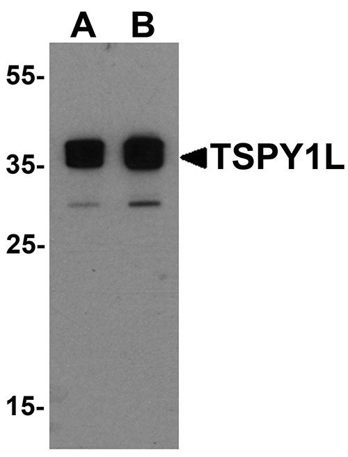
TSPY1L Antibody
- SPECIFICATION
- CITATIONS
- PROTOCOLS
- BACKGROUND

Application
| WB, E |
|---|---|
| Primary Accession | Q01534 |
| Other Accession | NP_003299, 139948460 |
| Reactivity | Human, Mouse |
| Host | Rabbit |
| Clonality | Polyclonal |
| Isotype | IgG |
| Calculated MW | 34 kDa |
| Application Notes | TSPY1L antibody can be used for detection of TSPY1L by Western blot at 0.5 - 1 µg/mL. |
| Gene ID | 7258 |
|---|---|
| Target/Specificity | TSPY1; TSPY1L antibody is human and mouse reactive. At least three isoforms of TSPY1 are known to exist; this antibody will detect only TSPY1L. |
| Reconstitution & Storage | TSPY1L antibody can be stored at 4℃ for three months and -20℃, stable for up to one year. As with all antibodies care should be taken to avoid repeated freeze thaw cycles. Antibodies should not be exposed to prolonged high temperatures. |
| Precautions | TSPY1L Antibody is for research use only and not for use in diagnostic or therapeutic procedures. |
| Name | TSPY1 |
|---|---|
| Synonyms | TSPY |
| Function | May be involved in sperm differentiation and proliferation. |
| Cellular Location | Cytoplasm. Nucleus. Note=Predominantly cytoplasmic. Also found in nucleus |
| Tissue Location | Specifically expressed in testicular tissues. Isoform 1 and isoform 2 are expressed in spermatogonia and spermatocytes. Found in early testicular carcinoma in situ, spermatogonial cells in testicular tissues of 46,X,Y female and in prostate cancer cell lines. |

Thousands of laboratories across the world have published research that depended on the performance of antibodies from Abcepta to advance their research. Check out links to articles that cite our products in major peer-reviewed journals, organized by research category.
info@abcepta.com, and receive a free "I Love Antibodies" mug.

Provided below are standard protocols that you may find useful for product applications.
Background
TSPY1L Antibody: Testis-specific protein on Y chromosome (TSPY1) is an ampliconic gene on the Y chromosome that has been associated with gonadoblastoma. Recent experiments have shown that in androgen-dependent testicular germ-cell tumors, TSPY1 can repress the androgen-bound androgen receptor (AR), a member of the nuclear steroid hormone receptor family that acts as a ligand-inducible transcription factor, suggesting that TSPY1 is a repressor of cell proliferation in germ-cell tumors and potentially in normal gonadal cells during early development. Two distinct isoforms of TSPY1, TSPY1L and TSPY1S, are known to exist.
References
Arnemann J, Jakubiczka S, Thuring S, et al. Cloning and sequence analysis of a human Y-chromosome-derived, testicular cDNA, TSPY. Genomics 1991; 11:108-114.
Lau YF. Gonadoblastoma, testicular and prostate cancers, and the TSPY gene. Am. J. Hum. Genet. 1999; 64:921-7.
Akimoto C, Ueda T, Inoue K, et al. Testis-specific protein on Y chromosome (TSPY) represses the activity of the androgen receptor in androgen-dependent testicular germ-cell tumors. Proc. Natl. Acad. Sci. USA 2010; 107:19891-6.
Krick R, Jacubiczka S, and Arnemann J. Expression, alternative splicing and haplotype analysis of transcribed testis specific protein (TSPY) genes. Gene 2003; 302:11-9.
If you have used an Abcepta product and would like to share how it has performed, please click on the "Submit Review" button and provide the requested information. Our staff will examine and post your review and contact you if needed.
If you have any additional inquiries please email technical services at tech@abcepta.com.
Ordering Information
Other Products
Shipping Information
