


 Foundational characteristics of cancer include proliferation, angiogenesis, migration, evasion of apoptosis, and cellular immortality. Find key markers for these cellular processes and antibodies to detect them.
 The SUMOplot™ Analysis Program predicts and scores sumoylation sites in your protein. SUMOylation is a post-translational modification involved in various cellular processes, such as nuclear-cytosolic transport, transcriptional regulation, apoptosis, protein stability, response to stress, and progression through the cell cycle.
 The Autophagy Receptor Motif Plotter predicts and scores autophagy receptor binding sites in your protein. Identifying proteins connected to this pathway is critical to understanding the role of autophagy in physiological as well as pathological processes such as development, differentiation, neurodegenerative diseases, stress, infection, and cancer.
CCL2 Antibody
- SPECIFICATION
- CITATIONS
- PROTOCOLS
- BACKGROUND

Application
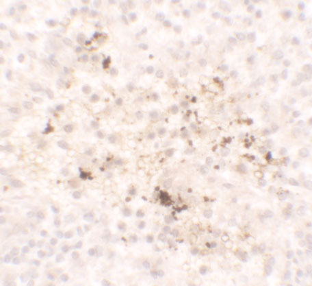
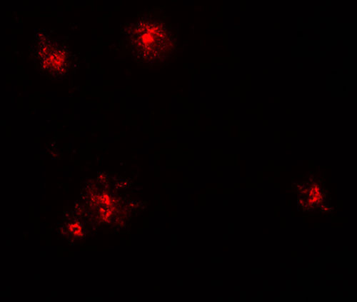
| WB, IHC-P, IF, E |
|---|---|
| Primary Accession | P13500 |
| Other Accession | NP_002973, 4506841 |
| Reactivity | Human, Mouse, Rat |
| Host | Rabbit |
| Clonality | Polyclonal |
| Isotype | IgG |
| Calculated MW | Predicted: 11 kDa |
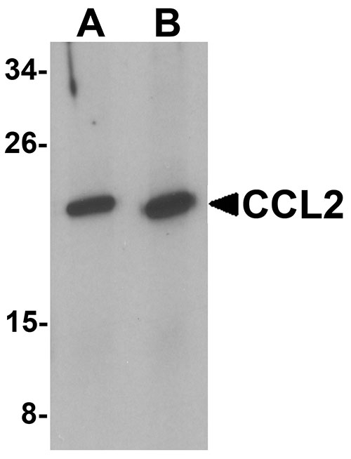
| Application Notes | CCL2 antibody can be used for detection of CCL2 by Western blot at 1 - 2 µg/mL. |
| Gene ID | 6347 |
|---|---|
| Target/Specificity | CCL2; |
| Reconstitution & Storage | CCL2 antibody can be stored at 4℃ for three months and -20℃, stable for up to one year. |
| Precautions | CCL2 Antibody is for research use only and not for use in diagnostic or therapeutic procedures. |
| Name | CCL2 |
|---|---|
| Synonyms | MCP1, SCYA2 |
| Function | Acts as a ligand for C-C chemokine receptor CCR2 (PubMed:10529171, PubMed:10587439, PubMed:9837883). Signals through binding and activation of CCR2 and induces a strong chemotactic response and mobilization of intracellular calcium ions (PubMed:10587439, PubMed:9837883). Exhibits a chemotactic activity for monocytes and basophils but not neutrophils or eosinophils (PubMed:8195247, PubMed:8627182, PubMed:9792674). May be involved in the recruitment of monocytes into the arterial wall during the disease process of atherosclerosis (PubMed:8107690). |
| Cellular Location | Secreted |
| Tissue Location | Expressed in the seminal plasma, endometrial fluid and follicular fluid (at protein level) (PubMed:23765988). Expressed in monocytes (PubMed:2513477). |

Thousands of laboratories across the world have published research that depended on the performance of antibodies from Abcepta to advance their research. Check out links to articles that cite our products in major peer-reviewed journals, organized by research category.
info@abcepta.com, and receive a free "I Love Antibodies" mug.

Provided below are standard protocols that you may find useful for product applications.
Background
CCL2 Antibody: CCL2, also known as monocyte chemotactic protein 1 (MCP1), belongs to the intercrine beta (chemokine CC) family. It is produced by a variety of cell types and is a potent chemoattractant for monocytes, memory T lymphocytes, and natural killer (NK) cells. It is upregulated during infection and inflammation. CCL2 is a potent basophil activator but does not affect eosinophils, whereas the related protein MCP2 stimulates both eosinophils and basophils. MCP3 has been shown to have the broadest range of influence. CCL2 has been implicated in the pathogenesis of diseases characterized by monocytic infiltrates, like psoriasis, rheumatoid arthritis or atherosclerosis.
References
Carr MW, Roth SJ, Luther E, et al. Monocyte chemoattractant protein 1 acts as a T-lymphocyte chemoattractant. Proc. Natl. Acad. Sci. USA 1994; 91:3652-6.
Conductier G, Blondeau N, Guyon A, et al. The role of monocyte chemoattractant protein MCP1/CCL2 in neuroinflammatory diseases. J. Neuroimmunol. 2010; 224:93-100.
Taub DD, Proost P, Murphy WJ, et al. Monocyte chemotactic protein-1 (MCP-1), -2, and -3 are chemotactic for human T lymphocytes. J. Clin. Invest. 1995; 95:1370-6
Bandinelli F, Del Rosso A, Gabrielli A, et al. CCL2, CCL3 and CCL5 chemokines in systemic sclerosis: the correlation with SSc clinical features and the effect of prostaglandin E1 treatment. Clin. Exp. Rheumatol. 2012; 30:S44-9.
If you have used an Abcepta product and would like to share how it has performed, please click on the "Submit Review" button and provide the requested information. Our staff will examine and post your review and contact you if needed.
If you have any additional inquiries please email technical services at tech@abcepta.com.
Ordering Information
Other Products
Shipping Information
