


 Foundational characteristics of cancer include proliferation, angiogenesis, migration, evasion of apoptosis, and cellular immortality. Find key markers for these cellular processes and antibodies to detect them.
 The SUMOplot™ Analysis Program predicts and scores sumoylation sites in your protein. SUMOylation is a post-translational modification involved in various cellular processes, such as nuclear-cytosolic transport, transcriptional regulation, apoptosis, protein stability, response to stress, and progression through the cell cycle.
 The Autophagy Receptor Motif Plotter predicts and scores autophagy receptor binding sites in your protein. Identifying proteins connected to this pathway is critical to understanding the role of autophagy in physiological as well as pathological processes such as development, differentiation, neurodegenerative diseases, stress, infection, and cancer.
CD59 Antibody
- SPECIFICATION
- CITATIONS
- PROTOCOLS
- BACKGROUND

Application
| WB, E |
|---|---|
| Primary Accession | P13987 |
| Other Accession | NP_000602, 10835165 |
| Reactivity | Human, Mouse |
| Host | Rabbit |
| Clonality | Polyclonal |
| Isotype | IgG |
| Calculated MW | 14 kDa |
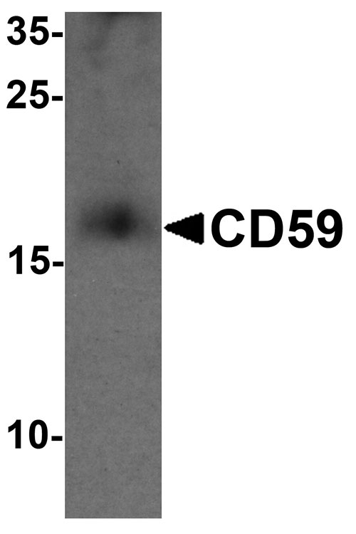
| Application Notes | CD59 antibody can be used for detection of CD59 by Western blot at 1 - 2 µg/mL. |
| Gene ID | 966 |
|---|---|
| Target/Specificity | CD59; |
| Reconstitution & Storage | CD59 antibody can be stored at 4℃ for three months and -20℃, stable for up to one year. |
| Precautions | CD59 Antibody is for research use only and not for use in diagnostic or therapeutic procedures. |
| Name | CD59 {ECO:0000303|PubMed:2475570, ECO:0000312|HGNC:HGNC:1689} |
|---|---|
| Function | Potent inhibitor of the complement membrane attack complex (MAC) action, which protects human cells from damage during complement activation (PubMed:11882685, PubMed:1698710, PubMed:2475111, PubMed:2475570, PubMed:2606909, PubMed:9053451). Acts by binding to the beta-haipins of C8 (C8A and C8B) components of the assembling MAC, forming an intermolecular beta-sheet that prevents incorporation of the multiple copies of C9 required for complete formation of the osmolytic pore (PubMed:11882685, PubMed:1698710, PubMed:36797260). |
| Cellular Location | Cell membrane; Lipid-anchor, GPI-anchor. Secreted. Note=Localizes to the cell surface (PubMed:36797260). Soluble form found in a number of tissues (PubMed:8670172). |

Thousands of laboratories across the world have published research that depended on the performance of antibodies from Abcepta to advance their research. Check out links to articles that cite our products in major peer-reviewed journals, organized by research category.
info@abcepta.com, and receive a free "I Love Antibodies" mug.

Provided below are standard protocols that you may find useful for product applications.
Background
CD59 Antibody: The complement regulatory protein CD59 is a cell surface glycoprotein that regulates complement-mediated cell lysis and is involved in lymphocyte signal transduction. CD59 is a potent inhibitor of the complement membrane attack complex, whereby it binds complement C8 and/or C9 during the assembly of this complex, thereby inhibiting the incorporation of multiple copies of C9 into the complex, which is necessary for osmolytic pore formation. CD59 also plays a role in signal transduction pathways in the activation of T cells. Mutations in this gene cause CD59 deficiency, a disease resulting in hemolytic anemia and thrombosis, and ultimately cerebral infarction.
References
Venneker GT and Asghar SS. CD59: a molecule involved in antigen presentation as well as downregulation of membrane attack complex. Exp. Clin. Immunogenet. 1992; 9:33-47.
Kimberly FC, Sivasankar B, and Paul Morgan B. Alternative roles for CD59. Mol. Immunol. 2007; 44:73-81.
Ninomiya H and Sims PJ. The human complement regulatory protein CD59 binds to the alpha-chain of C8 and to the “b” domain of C9. J. Biol. Chem. 1992; 267:13675-80.
Deckert M, Ticchioni M, Mari B, et al. The glycosylphosphatidylinositol-anchored CD59 protein stimulates both T cell receptor zeta/ZAP-70-dependent and –independent signaling pathways in T cells. Eur. J. Immunol. 1995; 25:1815-22
If you have used an Abcepta product and would like to share how it has performed, please click on the "Submit Review" button and provide the requested information. Our staff will examine and post your review and contact you if needed.
If you have any additional inquiries please email technical services at tech@abcepta.com.

Ordering Information
Other Products
Shipping Information
