


 Foundational characteristics of cancer include proliferation, angiogenesis, migration, evasion of apoptosis, and cellular immortality. Find key markers for these cellular processes and antibodies to detect them.
 The SUMOplot™ Analysis Program predicts and scores sumoylation sites in your protein. SUMOylation is a post-translational modification involved in various cellular processes, such as nuclear-cytosolic transport, transcriptional regulation, apoptosis, protein stability, response to stress, and progression through the cell cycle.
 The Autophagy Receptor Motif Plotter predicts and scores autophagy receptor binding sites in your protein. Identifying proteins connected to this pathway is critical to understanding the role of autophagy in physiological as well as pathological processes such as development, differentiation, neurodegenerative diseases, stress, infection, and cancer.
SOGA1 Antibody
- SPECIFICATION
- CITATIONS
- PROTOCOLS
- BACKGROUND

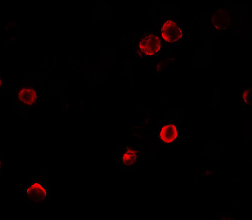
Application
| WB, IF, E |
|---|---|
| Primary Accession | O94964 |
| Other Accession | NP_954650, 66773344 |
| Reactivity | Human |
| Host | Rabbit |
| Clonality | Polyclonal |
| Isotype | IgG |
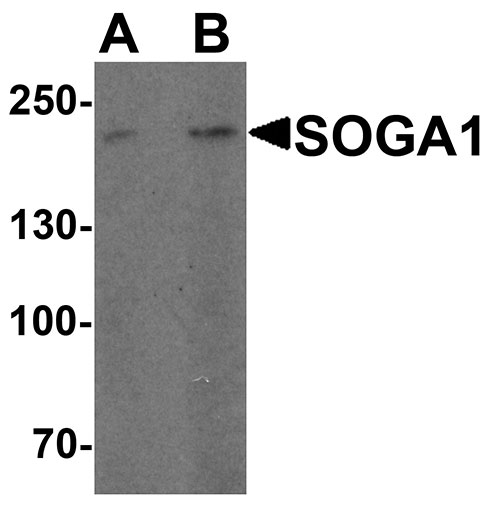
| Calculated MW | Predicted: 183 kDa Observed: 200kDa |
| Application Notes | SOGA1 antibody can be used for detection of SOGA1 by Western blot at 1 - 2 µg/ml. |
| Gene ID | 140710 |
|---|---|
| Target/Specificity | SOGA1; SOGA1 antibody is human specific. At least four isoforms of SOGA1 are known to exist. |
| Reconstitution & Storage | SOGA1 antibody can be stored at 4℃ for three months and -20℃, stable for up to one year. |
| Precautions | SOGA1 Antibody is for research use only and not for use in diagnostic or therapeutic procedures. |
| Name | MTCL2 (HGNC:16111) |
|---|---|
| Function | Microtubule-associated factor that enables integration of the centrosomal and Golgi-associated microtubules on the Golgi membrane, supporting directional migration. Preferentially acts on the perinuclear microtubules accumulated around the Golgi. Associates with the Golgi membrane through the N-terminal coiled-coil region and directly binds microtubules through the C-terminal domain (By similarity). Required for faithful chromosome segregation during mitosis (PubMed:33587225). Regulates autophagy by playing a role in the reduction of glucose production in an adiponectin- and insulin- dependent manner (By similarity). |
| Cellular Location | Cytoplasm, cytoskeleton. Golgi apparatus membrane {ECO:0000250|UniProtKB:E1U8D0}. Midbody Note=Associates with microtubules during late mitosis and interphase |

Thousands of laboratories across the world have published research that depended on the performance of antibodies from Abcepta to advance their research. Check out links to articles that cite our products in major peer-reviewed journals, organized by research category.
info@abcepta.com, and receive a free "I Love Antibodies" mug.

Provided below are standard protocols that you may find useful for product applications.
Background
The recently identified protein suppressor of glucose by autophagy protein 1 (SOGA1) has been found to be involved in the regulation of autophagy (1). SOGA1 is thought to contribute to adiponectin-mediated insulin-dependent inhibition of autophagy during the activation of adenosine monophosphate kinase (AMPK) (1,2). SOGA1 contains an internal signal peptide that enables the secretion of a circulating fragment of SOGA1, providing a surrogate marker for intracellular SOGA1 levels (2). Knockdown of SOGA1 elevated glucose production in primary hepatocytes indicates that SOGA1 is an inhibitor of glucose production. It thus might be useful as a novel therapeutic target for diabetes (3).
References
Cowherd RB, Asmar MM, Alderman JM, et al. Adiponectin lowers glucose production by increasing SOGA. Am. J. Pathol. 2010; 177:1936-45.
Madi T, Balamurugan K, Bombardi R, et al. The determination of tissue-specific DNA methylation patterns in forensic biofluids using bisulfite modification and pyrosequencing. Electrophoresis 2012; 33:1736-45.
Forbes JM. The physiological deadlock between AMPK and gluconeogenesis: SOGA, a novel protein, may provide the key. Am. J. Pathol. 2010; 177:1600-2.
If you have used an Abcepta product and would like to share how it has performed, please click on the "Submit Review" button and provide the requested information. Our staff will examine and post your review and contact you if needed.
If you have any additional inquiries please email technical services at tech@abcepta.com.
Ordering Information
Other Products
Shipping Information
