


 Foundational characteristics of cancer include proliferation, angiogenesis, migration, evasion of apoptosis, and cellular immortality. Find key markers for these cellular processes and antibodies to detect them.
 The SUMOplot™ Analysis Program predicts and scores sumoylation sites in your protein. SUMOylation is a post-translational modification involved in various cellular processes, such as nuclear-cytosolic transport, transcriptional regulation, apoptosis, protein stability, response to stress, and progression through the cell cycle.
 The Autophagy Receptor Motif Plotter predicts and scores autophagy receptor binding sites in your protein. Identifying proteins connected to this pathway is critical to understanding the role of autophagy in physiological as well as pathological processes such as development, differentiation, neurodegenerative diseases, stress, infection, and cancer.
UCP1 Antibody
- SPECIFICATION
- CITATIONS
- PROTOCOLS
- BACKGROUND

Application
| WB, IHC-P, E |
|---|---|
| Primary Accession | P25874 |
| Other Accession | NP_068605, 11225256 |
| Reactivity | Human, Mouse, Rat |
| Host | Rabbit |
| Clonality | Polyclonal |
| Isotype | IgG |
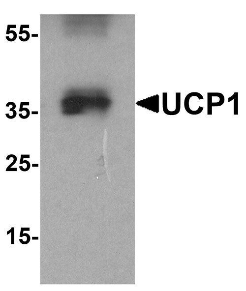
| Calculated MW | Predicted: 34 kDa Observed: 36 kDa |
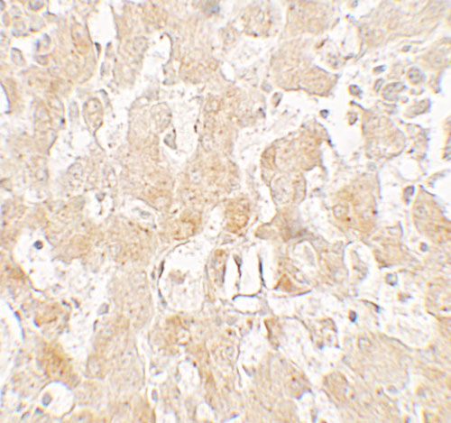
| Application Notes | UCP1 antibody can be used for detection of UCP1 by Western blot at 1 - 2 µg/ml. Antibody can also be used for Immunohistochemistry starting at 2.5 µg/mL. |
| Gene ID | 7350 |
|---|---|
| Target/Specificity | UCP1; UCP1 antibody is human, mouse and rat reactive. |
| Reconstitution & Storage | UCP1 antibody can be stored at 4℃ for three months and -20℃, stable for up to one year. |
| Precautions | UCP1 Antibody is for research use only and not for use in diagnostic or therapeutic procedures. |
| Name | UCP1 (HGNC:12517) |
|---|---|
| Function | Mitochondrial protein responsible for thermogenic respiration, a specialized capacity of brown adipose tissue and beige fat that participates in non-shivering adaptive thermogenesis to temperature and diet variations and more generally to the regulation of energy balance (By similarity). Functions as a long-chain fatty acid/LCFA and proton symporter, simultaneously transporting one LCFA and one proton through the inner mitochondrial membrane (PubMed:24196960, PubMed:28781081). However, LCFAs remaining associated with the transporter via their hydrophobic tails, it results in an apparent transport of protons activated by LCFAs. Thereby, dissipates the mitochondrial proton gradient and converts the energy of substrate oxydation into heat instead of ATP. Regulates the production of reactive oxygen species/ROS by mitochondria (By similarity). |
| Cellular Location | Mitochondrion inner membrane {ECO:0000250|UniProtKB:P12242}; Multi-pass membrane protein |
| Tissue Location | Brown adipose tissue.. |

Thousands of laboratories across the world have published research that depended on the performance of antibodies from Abcepta to advance their research. Check out links to articles that cite our products in major peer-reviewed journals, organized by research category.
info@abcepta.com, and receive a free "I Love Antibodies" mug.

Provided below are standard protocols that you may find useful for product applications.
Background
The mitochondrial brown fat uncoupling protein 1 (UCP1) is a member of the family of mitochondrial anion carrier proteins (MACP) (1). UCPs separate oxidative phosphorylation from ATP synthesis with energy dissipated as heat, also referred to as the mitochondrial proton leak. UCPs facilitate the transfer of anions from the inner to the outer mitochondrial membrane and the return transfer of protons from the outer to the inner mitochondrial membrane. They also reduce the mitochondrial membrane potential in mammalian cells (1). UCP1 is expressed only in brown adipose tissue, a specialized tissue which functions to produce heat (1).
References
1. Rial E, Gonzalez-Barroso MM, Fleury C, et al. The structure and function of the brown fat uncoupling protein UCP1: current status. Biofactors 1998; 8:209-19.
If you have used an Abcepta product and would like to share how it has performed, please click on the "Submit Review" button and provide the requested information. Our staff will examine and post your review and contact you if needed.
If you have any additional inquiries please email technical services at tech@abcepta.com.
Ordering Information
Other Products
Shipping Information
