


 Foundational characteristics of cancer include proliferation, angiogenesis, migration, evasion of apoptosis, and cellular immortality. Find key markers for these cellular processes and antibodies to detect them.
 The SUMOplot™ Analysis Program predicts and scores sumoylation sites in your protein. SUMOylation is a post-translational modification involved in various cellular processes, such as nuclear-cytosolic transport, transcriptional regulation, apoptosis, protein stability, response to stress, and progression through the cell cycle.
 The Autophagy Receptor Motif Plotter predicts and scores autophagy receptor binding sites in your protein. Identifying proteins connected to this pathway is critical to understanding the role of autophagy in physiological as well as pathological processes such as development, differentiation, neurodegenerative diseases, stress, infection, and cancer.
E2F3 Antibody
- SPECIFICATION
- CITATIONS
- PROTOCOLS
- BACKGROUND

Application
| WB, IHC-P, IF, E |
|---|---|
| Primary Accession | O00716 |
| Other Accession | NP_001940, 4503433 |
| Reactivity | Human, Mouse |
| Host | Rabbit |
| Clonality | Polyclonal |
| Isotype | IgG |
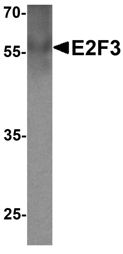
| Calculated MW | Predicted: 51 kDa Observed: 56 kDa |
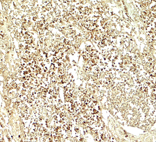
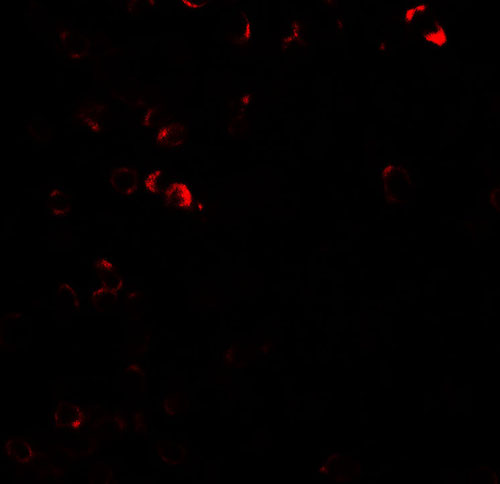
| Application Notes | E2F3 antibody can be used for detection of E2F3 by Western blot at 1 - 2 µg/ml. Antibody can also be used for Immunohistochemistry starting at 5 µg/mL. For immunofluorescence start at 20 µg/mL. |
| Gene ID | 1871 |
|---|---|
| Target/Specificity | E2F3; E2F3 antibody is human and mouse reactive. At least three isoforms of E2F3 are known to exist; this antibody will detect only the largest isoform. This antibody is predicted to not cross-react with other members of the E2F transcription factor family. |
| Reconstitution & Storage | E2F3 antibody can be stored at 4℃ for three months and -20℃, stable for up to one year. |
| Precautions | E2F3 Antibody is for research use only and not for use in diagnostic or therapeutic procedures. |
| Name | E2F3 |
|---|---|
| Synonyms | KIAA0075 |
| Function | Transcription activator that binds DNA cooperatively with DP proteins through the E2 recognition site, 5'-TTTC[CG]CGC-3' found in the promoter region of a number of genes whose products are involved in cell cycle regulation or in DNA replication. The DRTF1/E2F complex functions in the control of cell-cycle progression from G1 to S phase. E2F3 binds specifically to RB1 in a cell-cycle dependent manner. Inhibits adipogenesis, probably through the repression of CEBPA binding to its target gene promoters (By similarity). |
| Cellular Location | Nucleus. |

Thousands of laboratories across the world have published research that depended on the performance of antibodies from Abcepta to advance their research. Check out links to articles that cite our products in major peer-reviewed journals, organized by research category.
info@abcepta.com, and receive a free "I Love Antibodies" mug.

Provided below are standard protocols that you may find useful for product applications.
Background
The E2F transcription factor 3 (E2F3) is a member of a small family of transcription factors that function through binding of DP interaction partner proteins. E2F3 recognizes a specific sequence motif in DNA and interacts directly with the retinoblastoma protein (pRB) to regulate the expression of genes involved in the cell cycle (1). Like the related E2F1 and E2F2, E2F3 is essential for cellular proliferation and progression through the cell cycle (2). Altered copy number and activity of this gene have been observed in a number of human cancers (3).
References
Lees JA, Saito M, Vidal M, et al. The retinoblastoma protein binds to a family of E2F transcription factors. Mol. Cell. Biol. 1993; 13:7813-25.
Wu L, Timmers C, Maiti B, et al. The E2F1-3 transcription factors are essential for cellular proliferation. Nature 2001; 414:457-62.
DeGregori J. The genetics of the E2F family of transcription factors: shared functions and unique roles. Biochim. Biophys. Acta. 2002; 1602:131-50.
If you have used an Abcepta product and would like to share how it has performed, please click on the "Submit Review" button and provide the requested information. Our staff will examine and post your review and contact you if needed.
If you have any additional inquiries please email technical services at tech@abcepta.com.
Ordering Information
Other Products
Shipping Information
