


 Foundational characteristics of cancer include proliferation, angiogenesis, migration, evasion of apoptosis, and cellular immortality. Find key markers for these cellular processes and antibodies to detect them.
 The SUMOplot™ Analysis Program predicts and scores sumoylation sites in your protein. SUMOylation is a post-translational modification involved in various cellular processes, such as nuclear-cytosolic transport, transcriptional regulation, apoptosis, protein stability, response to stress, and progression through the cell cycle.
 The Autophagy Receptor Motif Plotter predicts and scores autophagy receptor binding sites in your protein. Identifying proteins connected to this pathway is critical to understanding the role of autophagy in physiological as well as pathological processes such as development, differentiation, neurodegenerative diseases, stress, infection, and cancer.
SPIB Antibody
- SPECIFICATION
- CITATIONS
- PROTOCOLS
- BACKGROUND

Application
| WB, IHC, E |
|---|---|
| Primary Accession | Q01892 |
| Other Accession | NP_003112, 61888836 |
| Reactivity | Human, Mouse, Rat |
| Host | Rabbit |
| Clonality | Polyclonal |
| Isotype | IgG |
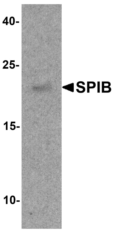
| Calculated MW | Predicted: 19, 25, 29 kDa; Observed: 20 kDa |
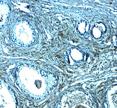
| Application Notes | SPIB antibody can be used for detection of SPIB by Western blot at 1 - 2 µg/ml. Antibody can also be used for immunohistochemistry starting at 2.5 µg/mL. |
| Gene ID | 6689 |
|---|---|
| Target/Specificity | SPIB; SPIB antibody is human, mouse and rat reactive. At least four isoforms of SPIB are known to exist; this antibody will detect all but isoform 3. |
| Reconstitution & Storage | SPIB antibody can be stored at 4℃ for three months and -20℃, stable for up to one year. |
| Precautions | SPIB Antibody is for research use only and not for use in diagnostic or therapeutic procedures. |
| Name | SPIB |
|---|---|
| Function | Sequence specific transcriptional activator which binds to the PU-box, a purine-rich DNA sequence (5'-GAGGAA-3') that can act as a lymphoid-specific enhancer. Promotes development of plasmacytoid dendritic cells (pDCs), also known as type 2 DC precursors (pre-DC2) or natural interferon (IFN)-producing cells. These cells have the capacity to produce large amounts of interferon and block viral replication. May be required for B-cell receptor (BCR) signaling, which is necessary for normal B-cell development and antigenic stimulation. |
| Cellular Location | [Isoform 1]: Nucleus |
| Tissue Location | Expressed in plasmacytoid dendritic cells (pDCs) and B-cells, not expressed in T-cells or granulocytes. May also be enriched in stem cell populations of the liver |

Thousands of laboratories across the world have published research that depended on the performance of antibodies from Abcepta to advance their research. Check out links to articles that cite our products in major peer-reviewed journals, organized by research category.
info@abcepta.com, and receive a free "I Love Antibodies" mug.

Provided below are standard protocols that you may find useful for product applications.
Background
SPIB is a member of the ETS transcription factor family that influences lymphoid development and activity and binds the consensus DNA site GGA (A/T) through a unique winged helix-turn-helix motif known as the ETS domain (1). SPIB is found in hematopoietic cells such as B cells and plasmacytoid dendritic cells (DC) (2,3). It promotes the development of plasmacytoid dendritic cells (pDCs) or natural interferon (IFN)-producing cells. SPIB may be required for B-cell receptor (BCR) signaling, which is necessary for normal B-cell development and antigenic stimulation (3,4).
References
Ray D, Bosselut R, Ghysdael J, et al. Characterization of Spi-B, a transcription factor related to the putative oncoprotein Spi-1/PU.1. Mol. Cell Biol. 1992; 12:4297-304.
Nagy M, Chapuis B, and Matthes T. Expression of transcription factors Pu.1, Spi-B, Blimp-1, BSAP and oct-2 in normal human plasma cells and in multiple myeloma cells. Br. J. Haematol. 2002; 116:429-35.
Schotte R, Rissoan MC, Bendriss-Vermare N, et al. The transcription factor Spi-B is expressed in plasmacytoid DC precursors and inhibits T-, B-, and NK-cell development. Blood 2003; 101:1015-23.
Schotte R, Nagasawa M, Weijer K, et al. The ETS transcription factor Spi-B is required for human plasmacytoid dendritic cell development. J. Exp. Med. 2004; 200:1503-9.
If you have used an Abcepta product and would like to share how it has performed, please click on the "Submit Review" button and provide the requested information. Our staff will examine and post your review and contact you if needed.
If you have any additional inquiries please email technical services at tech@abcepta.com.
Ordering Information
Shipping Information
