


 Foundational characteristics of cancer include proliferation, angiogenesis, migration, evasion of apoptosis, and cellular immortality. Find key markers for these cellular processes and antibodies to detect them.
 The SUMOplot™ Analysis Program predicts and scores sumoylation sites in your protein. SUMOylation is a post-translational modification involved in various cellular processes, such as nuclear-cytosolic transport, transcriptional regulation, apoptosis, protein stability, response to stress, and progression through the cell cycle.
 The Autophagy Receptor Motif Plotter predicts and scores autophagy receptor binding sites in your protein. Identifying proteins connected to this pathway is critical to understanding the role of autophagy in physiological as well as pathological processes such as development, differentiation, neurodegenerative diseases, stress, infection, and cancer.
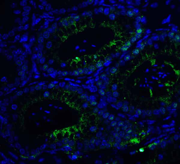
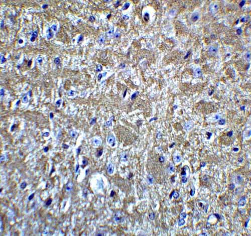
KIR2DS2 Antibody
- SPECIFICATION
- CITATIONS
- PROTOCOLS
- BACKGROUND

Application
| WB, IF, E |
|---|---|
| Primary Accession | P43631 |
| Other Accession | NP_036444, 100132285 |
| Reactivity | Human |
| Host | Rabbit |
| Clonality | Polyclonal |
| Isotype | IgG |
| Calculated MW | Predicted: 28, 33, 37 kDa Observed: 34, 42 kDa |
| Application Notes | KIR2DS2 antibody can be used for detection of KIR2DS2 by Western blot at 1 - 2 μg/ml. For immunofluorescence start at 20 μg/mL. |
| Gene ID | 100132285 |
|---|---|
| Target/Specificity | KIR2DS2 antibody was raised against an 18 amino acid peptide near the carboxy terminus of human KIR2DS2. The immunogen is located within the last 50 amino acids of KIR2DS2. |
| Reconstitution & Storage | Antibody can be stored at 4°C up to one year. Antibodies should not be exposed to prolonged high temperatures. |
| Precautions | KIR2DS2 Antibody is for research use only and not for use in diagnostic or therapeutic procedures. |
| Name | KIR2DS2 (HGNC:6334) |
|---|---|
| Synonyms | CD158J, NKAT5 |
| Function | Receptor on natural killer (NK) cells for HLA-C alleles. Does not inhibit the activity of NK cells. |
| Cellular Location | Cell membrane; Single-pass type I membrane protein |

Thousands of laboratories across the world have published research that depended on the performance of antibodies from Abcepta to advance their research. Check out links to articles that cite our products in major peer-reviewed journals, organized by research category.
info@abcepta.com, and receive a free "I Love Antibodies" mug.

Provided below are standard protocols that you may find useful for product applications.
Background
Killer cell immunoglobulin-like receptors (KIRs) are transmembrane glycoproteins expressed by natural killer cells and subsets of T cells (1). The KIR proteins are classified by the number of extracellular immunoglobulin domains and by whether they have a long (L) or short (S) cytoplasmic domain (2,3). KIR proteins are thought to play an important role in regulation of the immune response (3). KIR2DS2 downregulates the cytotoxicity of NK cells upon recognition of specific class I major histocompatibility complex (MHC) molecules on target cells and is a receptor on natural killer (NK) cells for HLA-C alleles (3,4).
References
Colonna M and Samaridis J. Cloning of immunoglobulin-superfamily members associated with HLA-C and HLA-B recognition by human natural killer cells. Science 1995; 268:405-8.
Biassoni R, Cantoni C, Falco M, et al. The human leukocyte antigen (HLA)-C-specific "activatory" or "inhibitory" natural killer cell receptors display highly homologous extracellular domains but differ in their transmembrane and intracytoplasmic portions. J. Exp. Med. 1996; 183:645-50.
Wagtmann N, Biassoni R, Cantoni C, et al. Molecular clones of the p58 NK cell receptor reveal immunoglobulin-related molecules with diversity in both the extra- and intracellular domains. Immunity 1995; 2:439-49.
Moesta AK and Parham P. Diverse functionality among human NK cell receptors for the C1 epitope of HLA-C: KIR2DS2, KIR2DL2, and KIR2DL3. Front. Immunol. 2012; 3:336.
If you have used an Abcepta product and would like to share how it has performed, please click on the "Submit Review" button and provide the requested information. Our staff will examine and post your review and contact you if needed.
If you have any additional inquiries please email technical services at tech@abcepta.com.
Ordering Information
Other Products
Shipping Information
