


 Foundational characteristics of cancer include proliferation, angiogenesis, migration, evasion of apoptosis, and cellular immortality. Find key markers for these cellular processes and antibodies to detect them.
 The SUMOplot™ Analysis Program predicts and scores sumoylation sites in your protein. SUMOylation is a post-translational modification involved in various cellular processes, such as nuclear-cytosolic transport, transcriptional regulation, apoptosis, protein stability, response to stress, and progression through the cell cycle.
 The Autophagy Receptor Motif Plotter predicts and scores autophagy receptor binding sites in your protein. Identifying proteins connected to this pathway is critical to understanding the role of autophagy in physiological as well as pathological processes such as development, differentiation, neurodegenerative diseases, stress, infection, and cancer.
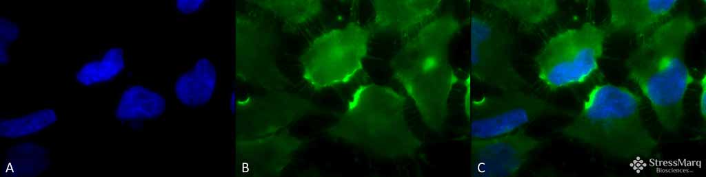
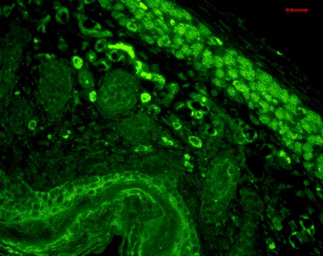
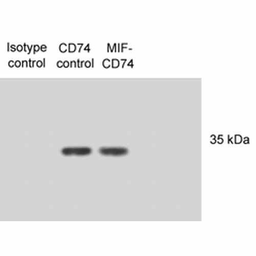
CD74 Antibody
CD74 Antibody, Clone PIN.1

- SPECIFICATION
- CITATIONS
- PROTOCOLS
- BACKGROUND

Application
| WB, IHC, ICC, IP, FC |
|---|---|
| Primary Accession | P04233 |
| Other Accession | NP_001020329.1 |
| Host | Mouse |
| Isotype | IgG |
| Reactivity | Human, Mouse |
| Clonality | Monoclonal |
| Description | Mouse Anti-Human CD74 Monoclonal IgG |
| Target/Specificity | Detects ~33-35kDa protein doublet corresponding to the molecular mass of the p33 and p35 forms of human CD74. |
| Other Names | DHLAG Antibody, HLA DR gamma Antibody, HLADG Antibody, p33 Antibody, p35 Antibody, Protein 41 Antibody, CD 74 antibody, CD74 antibody, CD74 antigen (invariant polypeptide of major histocompatibility complex, class II antigen-associated) antibody, CD74 antigen antibody, CD74 molecule antibody, CD74 molecule, major histocompatibility complex, class II invariant chain antibody, CLIP antibody, DHLAG antibody, Gamma chain of class II antigens antibody, HG2A_HUMAN antibody, HLA class II histocompatibility antigen gamma chain antibody, HLA DR antigens associated invariant chain antibody, HLA DR gamma antibody, HLA-DR antigens-associated invariant chain antibody, HLA-DR-gamma antibody, HLADG antibody, HLADR antigens associated invariant chain antibody Ia antigen associated invariant chain antibody, Ia antigen-associated invariant chain antibody, Ia associated invariant chain antibody, Ia gamma antibody, Ii antibody, Invariant polypeptide of major histocompatibility complex class II antigen associated antibody, la-gamma antibody, Major histocompatibility complex class II invariant chain antibody, MHC HLA DR gamma chain antibody, MHC HLA-DR gamma chain antibody, p33 antibody, p35 antibody, Protein 41 antibody |
| Clone Names | PIN.1 |
| Immunogen | Human CD74 invariant chain synthetic peptide |
| Purification | Protein G Purified |
| Storage | -20ºC |
| Storage Buffer | PBS pH7.2, 50% glycerol, 0.09% sodium azide |
| Shipping Temperature | Blue Ice or 4ºC |
| Certificate of Analysis | 1 µg/ml of SMC-116 was sufficient for detection of CD74 in 20 µg of PALA cell lysates by colorimetric immunolot analysis using goat anti-mouse IgG: AP as the secondary antibody. |
| Cellular Localization | Cell Membrane | Endoplasmic Reticulum | Endoplasmic Reticulum Membrane | Golgi Apparatus | Endosome | Lysosome |

Thousands of laboratories across the world have published research that depended on the performance of antibodies from Abcepta to advance their research. Check out links to articles that cite our products in major peer-reviewed journals, organized by research category.
info@abcepta.com, and receive a free "I Love Antibodies" mug.

Provided below are standard protocols that you may find useful for product applications.
Background
CD74 is a non-polymorphic type II integral membrane protein. It has a short N-terminal cytoplasmic tail of 28 amino acids, followed by a single 24-aa transmembrane region and an approximately 150-aa lumenal domain (1). The CD74 chain is thought to function mainly as an MHC class II chaperone, which promotes ER exit of MHC class II molecules, directs them to endocytic compartments, prevents peptide binding in the ER, and contributes to peptide editing in the MHC class II compartment. Class II MHC and Ii expression was believed to be restricted to classical antigen-presenting cells (APC); however, during inflammation, other cell types, including mucosal epithelial cells, have also been reported to express class II MHC molecules (2). Experiments that investigate cell-surface CD74 are complicated by the fact that CD74 remains on the cell surface for a very short time. The surface half-life of CD74 was calculated to be fewer than 10 minutes (3). CD74 however has also recently been shown to have a role as an accessory-signaling molecule because of its high-affinity binding to the pro-inflammatory cytokine, macrophage migration-inhibitory factor (MIF) (3). The restricted expression of CD74 by normal tissues and its very rapid internalization make CD74 an attractive therapeutic target for both cancer and immunologic diseases (4).
References
1. Becker-Hermann, S., Arie, G., Medvedovsky H, Kerem A, and Shachar I. (2005) Mol Bio Cell. 16(11):5061-9.
2. Barrera CA., et al (2005) J Histochem Cytochem 53 (12): 1481-9.
3. Starlets D., et al. (2006) Blood. 107 (12): 4807-4816.
4. Burton JD., et al. (2004). Clin Cancer Res. 10(19): 6606-11.
5. Denzin L.K., Hammond, C. and Cresswell, P. (1996) J. Exp. Med. 184: 2153-2165.
6. Denzin L.K., Robbins N.F., Carboy-Newcomb C. and Cresswell P. (1994) Immunity 1: 595-606.
If you have used an Abcepta product and would like to share how it has performed, please click on the "Submit Review" button and provide the requested information. Our staff will examine and post your review and contact you if needed.
If you have any additional inquiries please email technical services at tech@abcepta.com.
Ordering Information
Other Products
Shipping Information
