


 Foundational characteristics of cancer include proliferation, angiogenesis, migration, evasion of apoptosis, and cellular immortality. Find key markers for these cellular processes and antibodies to detect them.
 The SUMOplot™ Analysis Program predicts and scores sumoylation sites in your protein. SUMOylation is a post-translational modification involved in various cellular processes, such as nuclear-cytosolic transport, transcriptional regulation, apoptosis, protein stability, response to stress, and progression through the cell cycle.
 The Autophagy Receptor Motif Plotter predicts and scores autophagy receptor binding sites in your protein. Identifying proteins connected to this pathway is critical to understanding the role of autophagy in physiological as well as pathological processes such as development, differentiation, neurodegenerative diseases, stress, infection, and cancer.
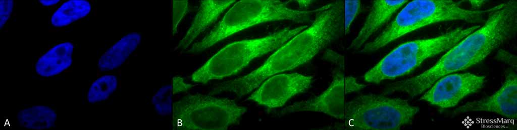
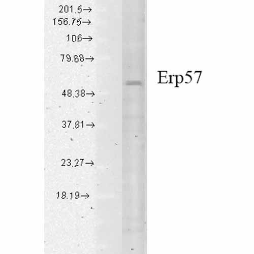
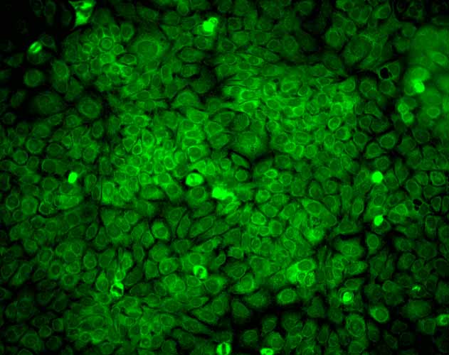
ERp57 (GRP58) Antibody
ERp57 (GRP58) Antibody, Clone Map.ERp57 (GRP58)

- SPECIFICATION
- CITATIONS
- PROTOCOLS
- BACKGROUND

Application
| WB, IHC, ICC, IP |
|---|---|
| Primary Accession | P30101 |
| Other Accession | NP_005304.3 |
| Host | Mouse |
| Isotype | IgG1 |
| Reactivity | Human, Mouse, Rat, Rabbit, Hamster, Monkey, Pig, Bovine, Guinea Pig, Dog |
| Clonality | Monoclonal |
| Description | Mouse Anti-Human ERp57 (GRP58) Monoclonal IgG1 |
| Target/Specificity | Detects ~57kDa |
| Other Names | ERp60 Antibody, ERp61 Antibody, Grp57 Antibody, Grp58 Antibody, P58 Antibody, PDIA3 Antibody, PI PLC Antibody, 58 kDa glucose regulated protein antibody, 58 kDa glucose-regulated protein antibody, 58 kDa microsomal protein antibody, Disulfide isomerase ER 60 antibody, Disulfide isomerase ER-60 antibody, Endoplasmic reticulum resident protein 57 antibody, Endoplasmic reticulum resident protein 60 antibody, ER p57 antibody, ER protein 57 antibody, ER protein 60 antibody, ERp 57 antibody, ERp57 antibody, Glucose Regulated Protein 58 Kd antibody, GRP 57 antibody, GRP 58 antibody, GRP57 antibody, HsT17083 antibody, p58 antibody, PDIA 3 antibody, PDIA3 antibody, PDIA3_HUMAN antibody, Phospholipase C alpha antibody, PI PLC antibody, Protein disulfide isomerase A3 antibody, Protein disulfide isomerase family A member 3 antibody, Protein disulfide-isomerase A3 antibody |
| Clone Names | Map.ERp57 |
| Immunogen | Human recombinant ERp57 (Grp58) |
| Purification | Protein G Purified |
| Storage | -20ºC |
| Storage Buffer | PBS pH7.4, 50% glycerol, 0.09% sodium azide |
| Shipping Temperature | Blue Ice or 4ºC |
| Certificate of Analysis | 0.5 µg/ml of SMC-168 was sufficient for detection of ERp57 in 10 µg of heat shock Heal Lysate by colorimetric immunoblot analysis using Goat anti-mouse IgG:HRP as the secondary antibody. |
| Cellular Localization | Endoplasmic Reticulum | Endoplasmic Reticulum Lumen | Melanosome |

Thousands of laboratories across the world have published research that depended on the performance of antibodies from Abcepta to advance their research. Check out links to articles that cite our products in major peer-reviewed journals, organized by research category.
info@abcepta.com, and receive a free "I Love Antibodies" mug.

Provided below are standard protocols that you may find useful for product applications.
Background
ERp57, also known as Glucose Regulated Protein 58 (Grp58), Hormone-Induced Protein-70 (HIP-70) and microsomal Carnitine Palmitoyltransferase, is a member of the protein disulfide isomerase family, containing two canonical CXHC tetrapeptide active site motifs (1-5). It has quite a few diverse roles. It functions as an accessory oxidoreductase involved in disulfide bond formation. In the ER, ERp57 interacts with membrane bound calnexin and soluble calreticulin (lectin chaperones) via their praline rich P-domain arms. Lectin chaperones bind nascent non-native glycoproteins, and position ERp57 to act upon the immature or misfolded glycoproteins that possess mono-glycosylated side chains. ERp57 deletion impairs posttranslational phases of influenza hema-glutinin folding, and causes accelerated release of MHC-I molecules, resulting in the coupling of sub-optimal peptides and reduced expression and stability on the cell surface (6). ERp57 also contains two thioredoxin active-site sequences, CGHC and an estrogen-binding domain. ERp57 is induced by both estrogen and leuteinizing-hormone-releasing hormone in the hippocampus (7).
References
1. Herbert D.N. and Molinari M. (2007) Physiol Rev. 87: 1377-1408.
2. Williams D.B. (2005) J Cell Sci. 119: 615-623
3. Maattanen P., et al. (2006) Biochem Cell Biol. 84: 881-889.
4. Oliver J.D., et al. (1999) Mol Bio Cell. 10: 2573-2582.
5. Oliver J.D., et al. (1997) Science 275: 86-88.
6. Solda T., et al. (2006) J Biol Chem 281: 6219-6226.
7. Kimura T., et al. (2005) Biochem Biophys Research Communications. 331 (1): 224-230.
8. Chen, G., et al. (2002) Clin Cancer Res 8(7): 2298-2305.
9. Tan, P., et al. F. (2002) J Immunol 168(4): 1950-1960.
If you have used an Abcepta product and would like to share how it has performed, please click on the "Submit Review" button and provide the requested information. Our staff will examine and post your review and contact you if needed.
If you have any additional inquiries please email technical services at tech@abcepta.com.
Ordering Information
Other Products
Shipping Information
