


 Foundational characteristics of cancer include proliferation, angiogenesis, migration, evasion of apoptosis, and cellular immortality. Find key markers for these cellular processes and antibodies to detect them.
 The SUMOplot™ Analysis Program predicts and scores sumoylation sites in your protein. SUMOylation is a post-translational modification involved in various cellular processes, such as nuclear-cytosolic transport, transcriptional regulation, apoptosis, protein stability, response to stress, and progression through the cell cycle.
 The Autophagy Receptor Motif Plotter predicts and scores autophagy receptor binding sites in your protein. Identifying proteins connected to this pathway is critical to understanding the role of autophagy in physiological as well as pathological processes such as development, differentiation, neurodegenerative diseases, stress, infection, and cancer.
PP5 Antibody
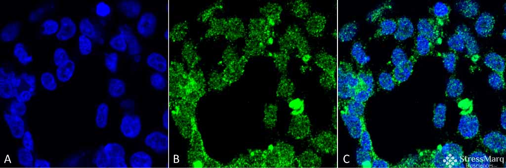
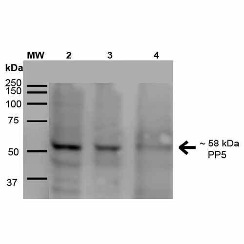
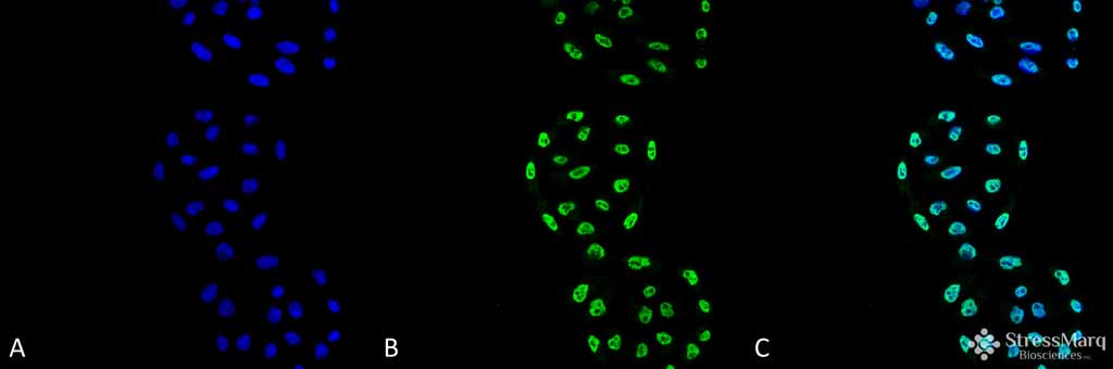
PP5 Antibody, Clone 2E11
- SPECIFICATION
- CITATIONS
- PROTOCOLS
- BACKGROUND

Application
| WB, ICC |
|---|---|
| Primary Accession | P53041 |
| Other Accession | NP_006238.1 |
| Host | Mouse |
| Isotype | IgG2a |
| Reactivity | Human |
| Clonality | Monoclonal |
| Description | Mouse Anti-Human PP5 Monoclonal IgG2a |
| Target/Specificity | Detects ~58kDa. |
| Other Names | PPP5 Antibody, PPT Antibody, Protein phosphatase T Antibody, Serine/threonine protein phosphatase 5 Antibody |
| Clone Names | 2E11 |
| Immunogen | Full length human PP5 protein |
| Purification | Protein G Purified |
| Storage | -20ºC |
| Storage Buffer | PBS pH7.4, 50% glycerol, 0.09% sodium azide |
| Shipping Temperature | Blue Ice or 4ºC |
| Certificate of Analysis | 1 µg/ml of SMC-243 was sufficient for detection of PP5 in 15 µg of human A431 lysates by ECL analysis using Goat anti-mouse IgG:HRP as the secondary antibody. |
| Cellular Localization | Nucleus | Cytoplasm |

Thousands of laboratories across the world have published research that depended on the performance of antibodies from Abcepta to advance their research. Check out links to articles that cite our products in major peer-reviewed journals, organized by research category.
info@abcepta.com, and receive a free "I Love Antibodies" mug.

Provided below are standard protocols that you may find useful for product applications.
Background
Proteins in this family participate in pathways regulated by reversible phosphorylation at serine and threonine residues, potentially playing a role in the regulation of cell growth and differentiation. PP5 or PPT is predominantly a nuclear protein and interacts with CDC16 and CDC27. It is found in association with several proteins that influence intracellular signlaing-cascades intitiated by hormones (glucocorticoids) or cellular stress (hypoxia, oxidative stress and DNA Damage). It phosphorylates serine residues of skeletal muscle phosphorylase and histone H1. It may also be involved in mitosis and RNA biogenesis regulation (1,2).
References
1. Butzow R., et al. (1988) J Lab Clin Med. 111(2): 249-256.
If you have used an Abcepta product and would like to share how it has performed, please click on the "Submit Review" button and provide the requested information. Our staff will examine and post your review and contact you if needed.
If you have any additional inquiries please email technical services at tech@abcepta.com.
Ordering Information
Other Products
Shipping Information
