


 Foundational characteristics of cancer include proliferation, angiogenesis, migration, evasion of apoptosis, and cellular immortality. Find key markers for these cellular processes and antibodies to detect them.
 The SUMOplot™ Analysis Program predicts and scores sumoylation sites in your protein. SUMOylation is a post-translational modification involved in various cellular processes, such as nuclear-cytosolic transport, transcriptional regulation, apoptosis, protein stability, response to stress, and progression through the cell cycle.
 The Autophagy Receptor Motif Plotter predicts and scores autophagy receptor binding sites in your protein. Identifying proteins connected to this pathway is critical to understanding the role of autophagy in physiological as well as pathological processes such as development, differentiation, neurodegenerative diseases, stress, infection, and cancer.
BVR Antibody
- SPECIFICATION
- CITATIONS
- PROTOCOLS
- BACKGROUND

Application
| WB, IHC, IP, ICC |
|---|---|
| Primary Accession | P53004 |
| Other Accession | NP_000703.2 |
| Host | Rabbit |
| Reactivity | Human |
| Clonality | Polyclonal |
| Description | Rabbit Anti-Human BVR Polyclonal |
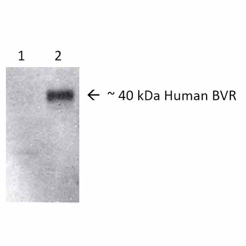
| Target/Specificity | Detects ~40-42kDa. |
| Other Names | Biliverdin Reductase Antibody, BIEA_HUMAN Antibody, Biliverdin IX alpha reductase Antibody, Biliverdin reductase A Antibody, Biliverdin-IX alpha-reductase Antibody, BLVR A Antibody, BLVR Antibody, Blvra Antibody, BVR A Antibody, BVRA Antibody, Zinc metalloprotein Antibody, zinc-metalloprotein Antibody |
| Immunogen | Human native full-length BVR |
| Purification | Protein A Purified |
| Storage | -20ºC |
| Storage Buffer | PBS pH7.4, 50% glycerol, 0.09% sodium azide |
| Shipping Temperature | Blue Ice or 4ºC |
| Certificate of Analysis | 1 µg/ml of SPC-214 was sufficient for detection of BVR in 10 µg of mixed human cell line lysate by colorimetric immunoblot analysis using Goat anti-rabbit IgG:HRP as the secondary antibody. |
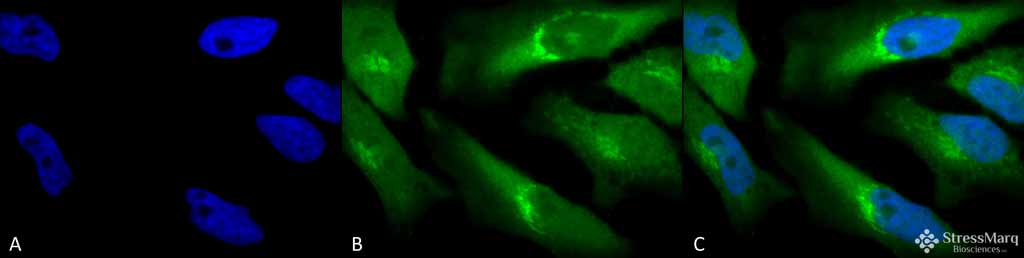
| Cellular Localization | Cytoplasm |

Thousands of laboratories across the world have published research that depended on the performance of antibodies from Abcepta to advance their research. Check out links to articles that cite our products in major peer-reviewed journals, organized by research category.
info@abcepta.com, and receive a free "I Love Antibodies" mug.

Provided below are standard protocols that you may find useful for product applications.
Background
Biliverdin Reductase (BVR) is a cytoplasmic enzyme that catalyzes the conversion of biliverdin to bilirubin by converting a double bond between the second and third pyrrole ring into a single bond (1). It is ubiqutiously expressed in all tissues- it occurs in cells and brain regiuons that already display HO-1 and HO-2, but also in regions and cell types with potential to induce stress proteins. It is unique among all enzymes in having two pH optima, using a different cofactor at each pH range, NADH at pH7.0 and NADPH at pH8.7 (2). It is not inactivated by heat shock, and have shown to abate inflammation, oxidative stress and apoptosis (3).
References
1. Singleton J.W., Laster L. (1965). J Biol Chem. 240: 4780-4789.
2. Kutty R.K., Maines M.D. (1981) J Biol Chem. 256: 3956-3962.
3. Mishra M., Ndisand J.F. (2014) Curr Pharm Des. 20(9): 1370-1391.
If you have used an Abcepta product and would like to share how it has performed, please click on the "Submit Review" button and provide the requested information. Our staff will examine and post your review and contact you if needed.
If you have any additional inquiries please email technical services at tech@abcepta.com.
Ordering Information
Other Products
Shipping Information
