


 Foundational characteristics of cancer include proliferation, angiogenesis, migration, evasion of apoptosis, and cellular immortality. Find key markers for these cellular processes and antibodies to detect them.
 The SUMOplot™ Analysis Program predicts and scores sumoylation sites in your protein. SUMOylation is a post-translational modification involved in various cellular processes, such as nuclear-cytosolic transport, transcriptional regulation, apoptosis, protein stability, response to stress, and progression through the cell cycle.
 The Autophagy Receptor Motif Plotter predicts and scores autophagy receptor binding sites in your protein. Identifying proteins connected to this pathway is critical to understanding the role of autophagy in physiological as well as pathological processes such as development, differentiation, neurodegenerative diseases, stress, infection, and cancer.
Alpha Synuclein Antibody (pSer129)
Rabbit Anti-Human Alpha Synuclein pSer129 Polyclonal
- SPECIFICATION
- CITATIONS
- PROTOCOLS
- BACKGROUND

Application
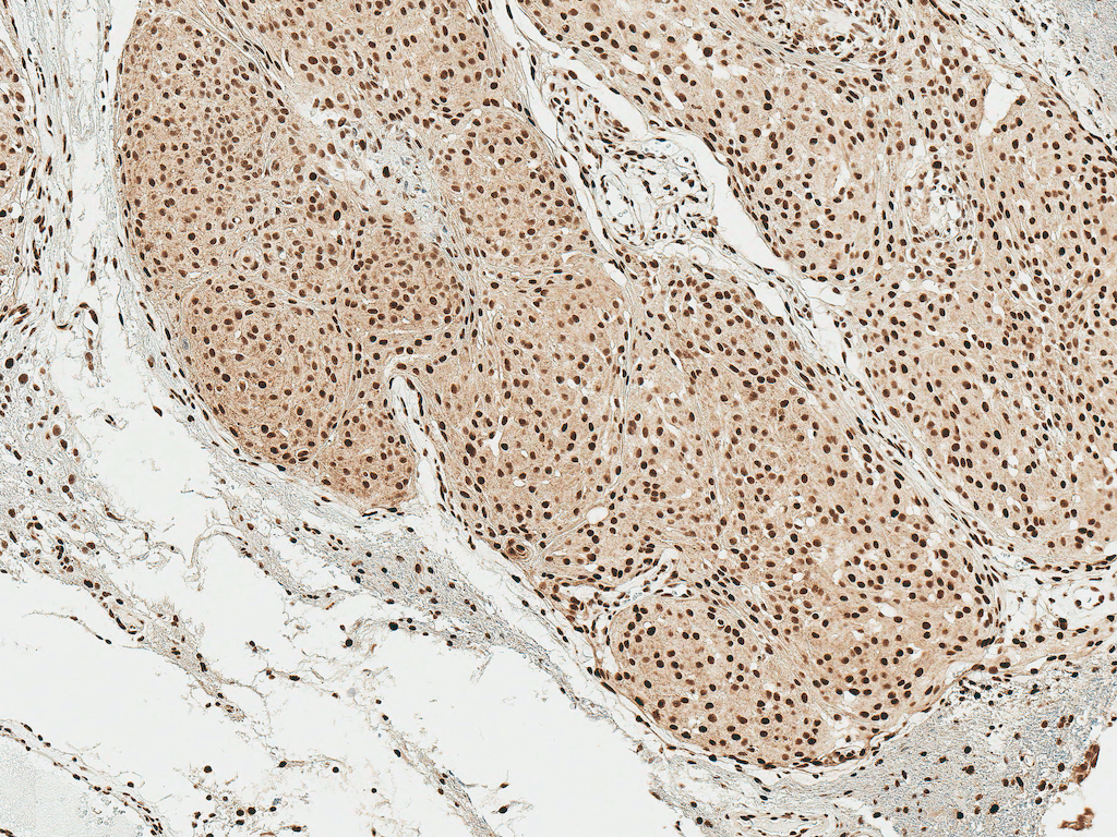
| IHC |
|---|---|
| Primary Accession | P37840 |
| Other Accession | NP_000336.1 |
| Host | Rabbit |
| Reactivity | Rat |
| Clonality | Polyclonal |
| Format | Alpha Synuclein (pSer129) |
| Target/Specificity | Alpha Synuclein (pSer129) |
| Other Names | Phospho anti-alpha Synuclein (S129) antibody, Alpha-synuclein (phospho S129) antibody, alpha Synuclein (phospho Ser129) antibody, alpha-Synuclein Phospho Ser129 Antibody, phospho-α-Synuclein (Ser129) Antibody, Alpha synuclein antibody, Alpha synuclein antibody phospho Serine 129, Alpha synuclein antibody phospho Ser 129, Alpha synuclein antibody pSerine 129, Alpha synuclein antibody pSer 129, Alpha synuclein antibody phosphoSer 129, Alpha-synuclein antibody, Alpha-synuclein, isoform NACP140 antibody, alphaSYN antibody, NACP antibody, Non A beta component of AD amyloid antibody, Non A4 component of amyloid antibody, Non A4 component of amyloid precursor antibody, Non-A beta component of AD amyloid antibody, Non-A-beta component of alzheimers disease amyloid , precursor of antibody, Non-A4 component of amyloid precursor antibody, Non-A4 component of amyloid, precursor of antibody, PARK 1 antibody, PARK 4 antibody, PARK1 antibody, PARK4 antibody, Parkinson disease (autosomal dominant, Lewy body) 4 antibody, Parkinson disease familial 1 antibody, SNCA antibody, alpha (non A4 component of amyloid precursor) antibody, SYN antibody, Synuclein alpha antibody, Synuclein alpha 140 antibody, Synuclein, alpha (non A4 component of amyloid precursor) antibody, SYUA_HUMAN antibody |
| Clone Names | Alpha Synuclein (pSer129) |
| Immunogen | Synthetic peptide of Human Alpha Synuclein pSer129 (40-140 aa), conjugated to Keyhole Limpet Haemocyanin (KLH). |
| Purification | Peptide Affinity Purified |
| Storage | -20ºC |
| Storage Buffer | PBS pH 7.4, 50% glycerol, 0.09% sodium azide *Storage buffer may change when conjugated |
| Shipping Temperature | Blue Ice or 4ºC |
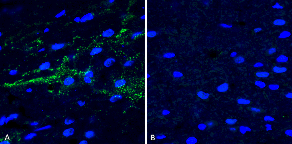
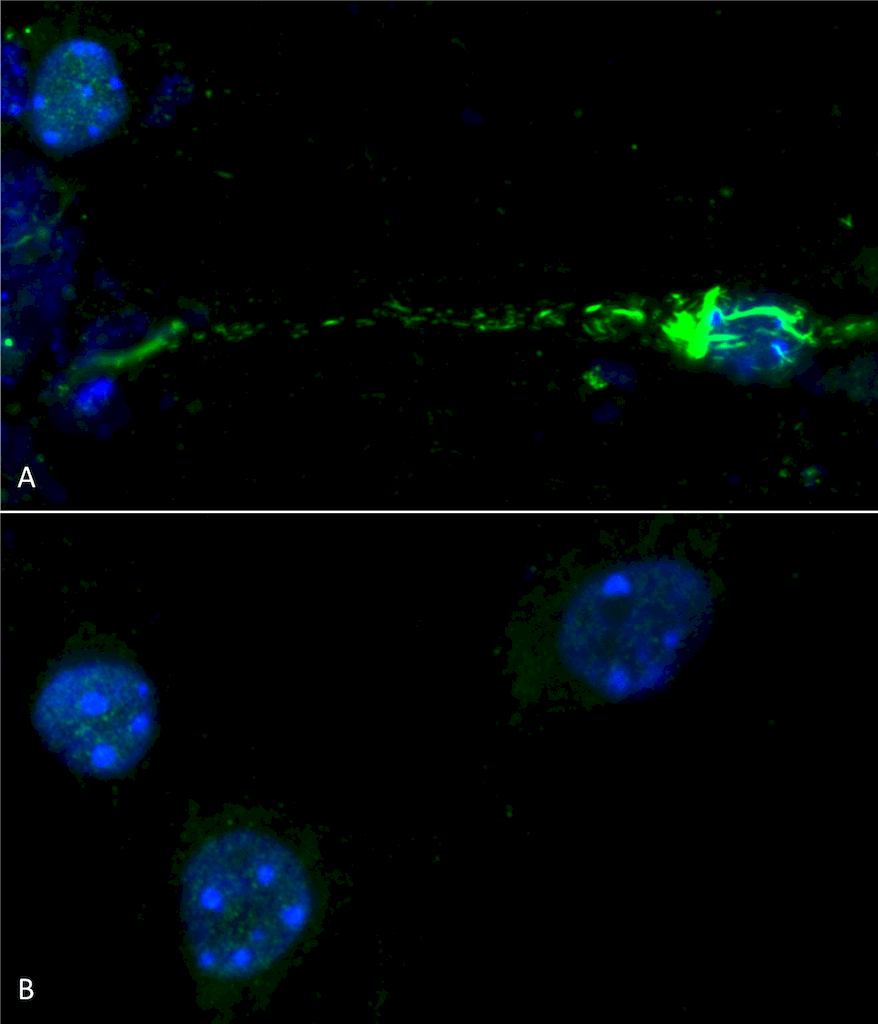
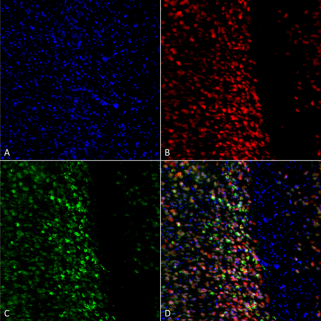
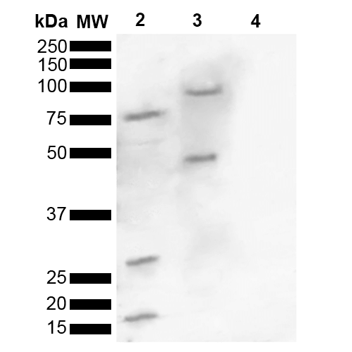
| Certificate of Analysis | A 1:1000 dilution of SPC-742 was sufficient for detection of Alpha Synuclein pSer129 in 15 µg of human brain cell lysates by ECL immunoblot analysis using goat anti-rabbit IgG:HRP as the secondary antibody. |
| Cellular Localization | Cytoplasm | Membrane | Nucleus | Cell Junction | Synapse | Secreted |

Thousands of laboratories across the world have published research that depended on the performance of antibodies from Abcepta to advance their research. Check out links to articles that cite our products in major peer-reviewed journals, organized by research category.
info@abcepta.com, and receive a free "I Love Antibodies" mug.

Provided below are standard protocols that you may find useful for product applications.
Background
Alpha-Synuclein (SNCA) is expressed predominantly in the brain, where it is concentrated in presynaptic nerve terminals (1). Alpha-synuclein is highly expressed in the mitochondria of the olfactory bulb, hippocampus, striatum and thalamus (2). Functionally, it has been shown to significantly interact with tubulin (3), and may serve as a potential microtubule-associated protein. It has also been found to be essential for normal development of the cognitive functions; inactivation may lead to impaired spatial learning and working memory (4). SNCA fibrillar aggregates represent the major non A-beta component of Alzheimers disease amyloid plaque, and a major component of Lewy body inclusions, and Parkinson's disease. Parkinson's disease (PD) is a common neurodegenerative disorder characterized by the progressive accumulation in selected neurons of protein inclusions containing alpha-synuclein and ubiquitin (5, 6). Alpha synuclein phosphorylated at serine 129 constitutes 90% of the alpha synuclein found in Lewy bodies (7, 8).
References
1. “Genetics Home Reference: SNCA”. US National Library of Medicine. (2013).
2. Zhang L., et al. (2008) Brain Res. 1244: 40-52.
3. Alim M.A., et al. (2002) J Biol Chem. 277(3): 2112-2117.
4. Kokhan V.S., Afanasyeva M.A., Van'kin G. (2012) Behav. Brain. Res. 231(1): 226-230.
5. Spillantini M.G., et al. (1997) Nature. 388(6645): 839-840.
6. Mezey E., et al. (1998) Nat Med. 4(7): 755-757.
7. Fujiwara H., et al. (2002) Nat Cell Biol. 4(2):160-4.
8. Anderson J.P., et al. (2006) J Biol Chem. 281(40):29739-52.
If you have used an Abcepta product and would like to share how it has performed, please click on the "Submit Review" button and provide the requested information. Our staff will examine and post your review and contact you if needed.
If you have any additional inquiries please email technical services at tech@abcepta.com.
Ordering Information
Other Products
Shipping Information
