


 Foundational characteristics of cancer include proliferation, angiogenesis, migration, evasion of apoptosis, and cellular immortality. Find key markers for these cellular processes and antibodies to detect them.
 The SUMOplot™ Analysis Program predicts and scores sumoylation sites in your protein. SUMOylation is a post-translational modification involved in various cellular processes, such as nuclear-cytosolic transport, transcriptional regulation, apoptosis, protein stability, response to stress, and progression through the cell cycle.
 The Autophagy Receptor Motif Plotter predicts and scores autophagy receptor binding sites in your protein. Identifying proteins connected to this pathway is critical to understanding the role of autophagy in physiological as well as pathological processes such as development, differentiation, neurodegenerative diseases, stress, infection, and cancer.
Tau Antibody
Rabbit Anti-Human Tau Polyclonal
- SPECIFICATION
- CITATIONS
- PROTOCOLS
- BACKGROUND

Application
| IHC |
|---|---|
| Primary Accession | P10636 |
| Host | Rabbit |
| Clonality | Polyclonal |
| Format | Tau |
| Target/Specificity | Tau |
| Other Names | Tau Antibody, Neurofibrillary tangle protein Antibody, MAPTL Antibody, Microtubule-associated protein tau Antibody, MTBT1 Antibody, Paired helical filament-tau Antibody, TAU Antibody, PHF-tau Antibody, MAPT Antibody, AI413597 antibody, AW045860 antibody, DDPAC antibody, FLJ31424 antibody, FTDP 17 antibody, G protein beta1/gamma2 subunit interacting factor 1 antibody, MAPT antibody, MAPTL antibody, MGC134287 antibody, MGC138549 antibody, MGC156663 antibody, Microtubule associated protein tau antibody, Microtubule associated protein tau isoform 4 antibody, Microtubule-associated protein tau antibody, MSTD antibody, Mtapt antibody, MTBT1 antibody, MTBT2 antibody, Neurofibrillary tangle protein antibody, Paired helical filament tau antibody, Paired helical filament-tau antibody, PHF tau antibody, PHF-tau antibody, PPND antibody, PPP1R103 antibody, Protein phosphatase 1, regulatory subunit 103 antibody, pTau antibody, RNPTAU antibody, TAU antibody, TAU_HUMAN antibody, Tauopathy and respiratory failure, included antibody |
| Clone Names | Tau |
| Immunogen | Recombinant Human Tau 2NR4 P301S Fibril |
| Purification | Protein A Purified |
| Storage | -20ºC |
| Storage Buffer | PBS pH 7.4, 50% glycerol, 0.09% sodium azide *Storage buffer may change when conjugated |
| Shipping Temperature | Blue Ice or 4ºC |
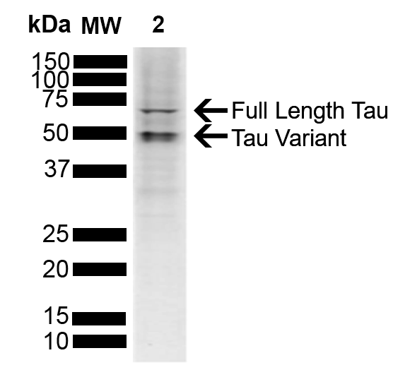
| Certificate of Analysis | A 1:1000 dilution of SPC-801 was sufficient for detection of Tau in 10 µg of human SH-SY5Y cell lysate cell lysates by ECL immunoblot analysis using goat anti-rabbit IgG:HRP as the secondary antibody. |
| Cellular Localization | Cytoskeleton | Plasma Membrane | Cell Membrane | Peripheral Membrane Protein | Cytoplasmic Side | Cytosol | Axon | Microtubule | Microtubule Associated Complex | Tubulin Complex | Nucleus | Nuclear Periphery | Nuclear Speck | Axolemma Plasma Membrane | Axolemma | Cytoplasm | Cell Body | Cytoplasmic Ribonucleoprotein Granule | Dendrite | Growth Cone | Neurofibrillary Tangle | Neuronal Cell Body |

Thousands of laboratories across the world have published research that depended on the performance of antibodies from Abcepta to advance their research. Check out links to articles that cite our products in major peer-reviewed journals, organized by research category.
info@abcepta.com, and receive a free "I Love Antibodies" mug.

Provided below are standard protocols that you may find useful for product applications.
Background
Alzheimer’s Disease (AD) is the most common neurodegenerative disease, affecting 10% of seniors over the age of 65 (1). It was named after Alois Alzheimer, a German scientist who discovered tangled bundles of fibrils where neurons had once been in the brain of a deceased patient in 1907 (2). Tau (tubulin-associated unit) is normally located in the axons of neurons where it stabilizes microtubules. Tauopathies such as AD are characterized by neurofibrillary tangles containing hyperphosphorylated tau fibrils (3). There are six isoforms of tau in the adult human brain: three with four repeat units (4R) and three with three repeat units (3R) (4). K18 is a truncated form of human tau containing only the 4 microtubule binding repeats (5). P301L (PL) is a mutation where proline is replaced by leucine at codon 301 of tau, and has been linked to frontotemporal dementia (6).
References
1. www.alz.org/alzheimers-dementia/facts-figures
2. Alzheimer, A. Über eine eigenartige Erkrankung der Hirnrinde. Allg. Z. Psychiatr. Psych.-Gerichtl. Med. 64, 146–148 (1907)
3. Matsumoto, G. et al. (2018). Int J Mol Sci. 19, 1497.
4. Goedert, M. and Spillantini, M. G. (2017). Mol Brain. 10:18.
5. Guo, J. and Lee, M.Y. (2013). FEBS Lett. 587(6): 717-723.
6. Alberici, A. et al. (2004). J Neurol Neurosurg Psychiatry. 75:1607-1610.
If you have used an Abcepta product and would like to share how it has performed, please click on the "Submit Review" button and provide the requested information. Our staff will examine and post your review and contact you if needed.
If you have any additional inquiries please email technical services at tech@abcepta.com.
Ordering Information
Other Products
Shipping Information
