


 Foundational characteristics of cancer include proliferation, angiogenesis, migration, evasion of apoptosis, and cellular immortality. Find key markers for these cellular processes and antibodies to detect them.
 The SUMOplot™ Analysis Program predicts and scores sumoylation sites in your protein. SUMOylation is a post-translational modification involved in various cellular processes, such as nuclear-cytosolic transport, transcriptional regulation, apoptosis, protein stability, response to stress, and progression through the cell cycle.
 The Autophagy Receptor Motif Plotter predicts and scores autophagy receptor binding sites in your protein. Identifying proteins connected to this pathway is critical to understanding the role of autophagy in physiological as well as pathological processes such as development, differentiation, neurodegenerative diseases, stress, infection, and cancer.
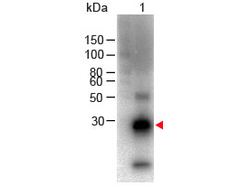
F(ab')2 Anti-Rabbit IgG F(c) (Biotin Conjugated) Secondary Antibody
Goat Polyclonal, Biotin
- SPECIFICATION
- CITATIONS
- PROTOCOLS
- BACKGROUND
| Description | F(ab')2 Anti-RABBIT IgG F(c) (GOAT) Antibody Biotin Conjugated |
|---|---|
| Host | Goat |
| Conjugate | Biotin |
| Target Species | Rabbit |
| Clonality | Polyclonal |
Application
| WB, E, IC |
| Application Note | ELISA 1:20,000-1:100,000 Western Blot 1:2,000-1:10,000 Immunochemistry 1:1,000-1:5,000 |
| Physical State | Lyophilized |
| Host Isotype | IgG F(ab')2 |
| Target Isotype | IgG F(c) |
| Buffer | 0.02 M Potassium Phosphate, 0.15 M Sodium Chloride, pH 7.2 |
| Immunogen | Rabbit IgG F(c) fragment |
| Reconstitution Volume | 500 µL |
| Reconstitution Buffer | Restore with deionized water (or equivalent) |
| Stabilizer | 10 mg/mL Bovine Serum Albumin (BSA) - Immunoglobulin and Protease free |
| Preservative | 0.01% (w/v) Sodium Azide |
| Shipping Condition | Ambient |
|---|---|
| Purity | F(ab') fragment biotin conjugated secondary antibody was prepared from monospecific antiserum by immunoaffinity chromatography using Rabbit IgG coupled to agarose beads followed by solid phase adsorption(s) to remove any unwanted reactivities, pepsin digestion and chromatographic separation. Assay by immunoelectrophoresis resulted in a single precipitin arc against anti-Biotin, anti-Goat Serum, Rabbit IgG, Rabbit IgG F(c) and Rabbit Serum. No reaction was observed against anti-Pepsin, anti-Goat IgG F(c) or Rabbit IgG F(ab’)2. |
| Storage Condition | Store vial at 4° C prior to restoration. For extended storage aliquot contents and freeze at -20° C or below. Avoid cycles of freezing and thawing. Centrifuge product if not completely clear after standing at room temperature. This product is stable for several weeks at 4° C as an undiluted liquid. Dilute only prior to immediate use. |
| Precautions Note | This product is for research use only and is not intended for therapeutic or diagnostic applications. |

Thousands of laboratories across the world have published research that depended on the performance of antibodies from Abcepta to advance their research. Check out links to articles that cite our products in major peer-reviewed journals, organized by research category.
info@abcepta.com, and receive a free "I Love Antibodies" mug.

Provided below are standard protocols that you may find useful for product applications.
Background
Suitable for immunomicroscopy and flow cytometry or FACS analysis as well as other antibody based fluorescent assays requiring lot-to-lot consistency.
If you have used an Abcepta product and would like to share how it has performed, please click on the "Submit Review" button and provide the requested information. Our staff will examine and post your review and contact you if needed.
If you have any additional inquiries please email technical services at tech@abcepta.com.
Ordering Information
Shipping Information
