


 Foundational characteristics of cancer include proliferation, angiogenesis, migration, evasion of apoptosis, and cellular immortality. Find key markers for these cellular processes and antibodies to detect them.
 The SUMOplot™ Analysis Program predicts and scores sumoylation sites in your protein. SUMOylation is a post-translational modification involved in various cellular processes, such as nuclear-cytosolic transport, transcriptional regulation, apoptosis, protein stability, response to stress, and progression through the cell cycle.
 The Autophagy Receptor Motif Plotter predicts and scores autophagy receptor binding sites in your protein. Identifying proteins connected to this pathway is critical to understanding the role of autophagy in physiological as well as pathological processes such as development, differentiation, neurodegenerative diseases, stress, infection, and cancer.
Anti-TRPC6 (MOUSE) Monoclonal Antibody
TRPC6 Antibody
- SPECIFICATION
- CITATIONS
- PROTOCOLS
- BACKGROUND

| Host | Mouse |
|---|---|
| Conjugate | Unconjugated |
| Target Species | Human |
| Reactivity | Human, Mouse |
| Clonality | Monoclonal |
Application
| WB, IHC, E, I, LCI |
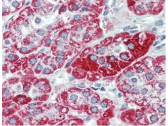
| Application Note | Anti-TRPC6 monoclonal antibody has been tested by ELISA, immunohistochemistry and western blotting. Expect a band approximately 30 kDa in size corresponding to the cytoplasmic domain of TRPC6 protein by western blotting in the appropriate cell lysate or extract. Specific conditions for reactivity should be optimized by the end user. Use formalin-fixed paraffin-embedded sections for immunohistochemistry. No pre-treatment of sample is required. Strong staining was observed in adrenal, Purkinje neurons, cortical neurons, heart, ganglion cells, renal tubules, Sertoli cells, hepatocytes, skeletal muscle, exocrine pancreas, and germinal centers of lymphoid follicles. Moderate staining was observed in colon epithelium and plasma cells, B-lymphocytes, and parafollicular cells of the thyroid. Faint staining was seen in respiratory epithelium. Prostate and placenta were negative for staining. The antibody produced minimal to no background staining and appeared very specific at 2.5 µg/mL. |
| Physical State | Liquid (sterile filtered) |
| Buffer | 0.02 M Potassium Phosphate, 0.15 M Sodium Chloride, pH 7.2 |
| Immunogen | This monoclonal antibody was produced by repeated immunizations with a synthetic peptide corresponding to a region near the carboxy terminus of human TRPC6 protein. |
| Preservative | 0.01% (w/v) Sodium Azide |
| Gene ID | 7225 |
|---|---|
| Other Names | 7225 |
| Purity | This product was purified from concentrated tissue culture supernate by Protein A chromatography. This antibody is specific for human TRPC6 protein. A BLAST analysis was used to suggest cross-reactivity with TRPC6 from chimpanzee based on 100% homology with the immunizing sequence. Cross-reactivity with TRPC6 from other sources has not been determined. |
| Storage Condition | Store vial at -20° C prior to opening. Aliquot contents and freeze at -20° C or below for extended storage. Avoid cycles of freezing and thawing. Centrifuge product if not completely clear after standing at room temperature. This product is stable for several weeks at 4° C as an undiluted liquid. Dilute only prior to immediate use. |
| Precautions Note | This product is for research use only and is not intended for therapeutic or diagnostic applications. |
| Name | TRPC6 {ECO:0000303|PubMed:9930701, ECO:0000312|HGNC:HGNC:12338} |
|---|---|
| Function | Forms a receptor-activated non-selective calcium permeant cation channel (PubMed:19936226, PubMed:23291369, PubMed:26892346, PubMed:9930701). Probably is operated by a phosphatidylinositol second messenger system activated by receptor tyrosine kinases or G-protein coupled receptors. Activated by diacylglycerol (DAG) in a membrane- delimited fashion, independently of protein kinase C (PubMed:26892346). Seems not to be activated by intracellular calcium store depletion. |
| Cellular Location | Cell membrane; Multi-pass membrane protein |
| Tissue Location | Expressed primarily in placenta, lung, spleen, ovary and small intestine. Expressed in podocytes and is a component of the glomerular slit diaphragm. |

Thousands of laboratories across the world have published research that depended on the performance of antibodies from Abcepta to advance their research. Check out links to articles that cite our products in major peer-reviewed journals, organized by research category.
info@abcepta.com, and receive a free "I Love Antibodies" mug.

Provided below are standard protocols that you may find useful for product applications.
Background
TRPC6, also known as TRP6, short transient receptor potential channel 6 and transient receptor potential cation channel subfamily C member 6, is thought to form a receptor-activated non-selective calcium permeant cation channel. TRPC6 is probably operated by a phosphatidylinositol second messenger system activated by receptor tyrosine kinases or G-protein coupled receptors. It is activated by diacylglycerol (DAG) in a membrane-delimited fashion, independently of protein kinase C and may not to be activated by intracellular calcium store depletion. Defects in this gene are a cause of focal segmental glomerulosclerosis (FSGS). Expression of this protein has been reported in tissues such as placenta, lung, spleen, ovary, small intestine, and renal podocytes. Immunohistochemistry studies using polyclonal antibodies to this target have shown moderate to strong staining in cell types such as neurons, breast, respiratory, squamous and prostate epithelium, epidermis, placental trophoblasts, dendritic cells, and subsets of immune cells, and faint to moderate staining of adrenal, colon, ganglion cells, hepatocytes, heart, and testis.
If you have used an Abcepta product and would like to share how it has performed, please click on the "Submit Review" button and provide the requested information. Our staff will examine and post your review and contact you if needed.
If you have any additional inquiries please email technical services at tech@abcepta.com.
Ordering Information
Other Products
Shipping Information
