


 Foundational characteristics of cancer include proliferation, angiogenesis, migration, evasion of apoptosis, and cellular immortality. Find key markers for these cellular processes and antibodies to detect them.
 The SUMOplot™ Analysis Program predicts and scores sumoylation sites in your protein. SUMOylation is a post-translational modification involved in various cellular processes, such as nuclear-cytosolic transport, transcriptional regulation, apoptosis, protein stability, response to stress, and progression through the cell cycle.
 The Autophagy Receptor Motif Plotter predicts and scores autophagy receptor binding sites in your protein. Identifying proteins connected to this pathway is critical to understanding the role of autophagy in physiological as well as pathological processes such as development, differentiation, neurodegenerative diseases, stress, infection, and cancer.
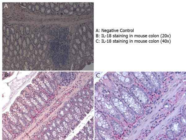
Anti-Mouse IL-18 (RABBIT) Antibody
Mouse IL-18 Antibody
- SPECIFICATION
- CITATIONS
- PROTOCOLS
- BACKGROUND

| Host | Rabbit |
|---|---|
| Conjugate | Unconjugated |
| Target Species | Mouse |
| Reactivity | Mouse |
| Clonality | Polyclonal |
Application
| WB, IHC, E, I, LCI |
| Application Note | Anti-Mouse IL-18 has been tested in immunohistochemistry and immunofluorescence and is suitable for use in neutralizations, ELISA, and immunoblotting. Although untested, this reagent may be useful for radioimmunoassays, flow cytometry and immunoprecipitation. It recognizes the 18,000 MW mature (active) IL-18. Reactivity in other immunoassays is unknown. |
| Physical State | Liquid (sterile filtered) |
| Buffer | 0.02 M Potassium Phosphate, 0.15 M Sodium Chloride, pH 7.2 |
| Immunogen | The whole rabbit serum used to produce this IgG fraction antibody was prepared by repeated immunizations with native 157 aa mouse IL-18 produced in E.coli. |
| Gene ID | 16173 |
|---|---|
| Other Names | 16173 |
| Purity | This is an IgG preparation of whole rabbit serum purified by protein A chromatography using a low endotoxin methodology. This antibody is primarily directed against mature 18,000 MW mouse IL-18 and is useful in determining its presence in various assays. This antibody will also recognize the 24,000 inactive precursor form of mouse IL-18. In general, this antibody also detects rat IL-18 in the same formats using similar dilutions. A control of similarly diluted LOW ENDOTOXIN CONTROL RABBIT IgG (code # 011-001-297) is recommended. |
| Storage Condition | Store vial at -20° C prior to opening. Aliquot contents and freeze at -20° C or below for extended storage. Avoid cycles of freezing and thawing. Centrifuge product if not completely clear after standing at room temperature. This product is stable for several weeks at 4° C as an undiluted liquid. Dilute only prior to immediate use. |
| Precautions Note | This product is for research use only and is not intended for therapeutic or diagnostic applications. |
| Name | Il18 {ECO:0000312|MGI:MGI:107936} |
|---|---|
| Synonyms | Igif |
| Function | Pro-inflammatory cytokine primarily involved in epithelial barrier repair, polarized T-helper 1 (Th1) cell and natural killer (NK) cell immune responses (PubMed:26638072, PubMed:26638073). Upon binding to IL18R1 and IL18RAP, forms a signaling ternary complex which activates NF-kappa-B, triggering synthesis of inflammatory mediators (By similarity). Synergizes with IL12/interleukin-12 to induce IFNG synthesis from T-helper 1 (Th1) cells and natural killer (NK) cells (By similarity). Involved in transduction of inflammation downstream of pyroptosis: its mature form is specifically released in the extracellular milieu by passing through the gasdermin-D (GSDMD) pore (PubMed:30392956). |
| Cellular Location | Cytoplasm {ECO:0000250|UniProtKB:Q14116}. Secreted. Note=The precursor is cytosolic (By similarity). In response to inflammasome-activating signals, cleaved and secreted (By similarity). Mature form is secreted and released in the extracellular milieu by passing through the gasdermin-D (GSDMD) pore (PubMed:30392956). In contrast, the precursor form is not released, due to the presence of an acidic region that is proteolytically removed by CASP1 during maturation (By similarity). The secretion is dependent on protein unfolding and facilitated by the cargo receptor TMED10 (By similarity). {ECO:0000250|UniProtKB:Q14116, ECO:0000269|PubMed:30392956} |

Thousands of laboratories across the world have published research that depended on the performance of antibodies from Abcepta to advance their research. Check out links to articles that cite our products in major peer-reviewed journals, organized by research category.
info@abcepta.com, and receive a free "I Love Antibodies" mug.

Provided below are standard protocols that you may find useful for product applications.
Background
Interleukin-18 (IL-18) is a member of the IL-1 cytokine family and was initially identified as an Interferon-g (IFN-g) inducing factor (IGIF). The IL-18 gene was originally cloned from liver cells and has since been shown to be produced by activated monocytes/ macrophages, Kupffer cells, keratinocytes, glucocorticoid-secreting adrenal cortex cells, osteoblasts and dendritic cells. IL-18 is a 24 kDa, non-glycosylated polypeptide that lacks a classical signal sequence and possesses a structure recognizably similar to IL-1. IL-18 is synthesized as a bio-inactive propeptide that undergoes proteolytic cleavage by either ICE (interleukin-1 beta converting enzyme) or another caspase to generate a mature, bioactive, 18 kDa molecule. In both the mature and propeptide forms, IL-18 shows 64% aa sequence identity from mouse to human. IL-18 does not appear to show any primary sequence similarity to any other known cytokines. Rat IL-18 has also been isolated, and found to be 194 aa in length with a 91% aa sequence identity to mouse IL-18. Human IL-18 has been found to induce the production of IFN-g and GM-CSF while inhibiting the production of IL-10 by PBMC. With respect to human T cells, IL-18 enhances Th1 cytokine production and stimulates cell proliferation via an IL-2-dependent pathway. Human IL-18 can also inhibit the synthesis of IgE by B cells. Thus, IL-18 plays an important role in immunological and inflammatory reactions. Currently, the bioactivity of human IL-18 is often determined by its capacity to augment the levels of IFN-g produced by T cells as measured in tissue culture supernatants.
If you have used an Abcepta product and would like to share how it has performed, please click on the "Submit Review" button and provide the requested information. Our staff will examine and post your review and contact you if needed.
If you have any additional inquiries please email technical services at tech@abcepta.com.
Ordering Information
Other Products
Shipping Information
