


 Foundational characteristics of cancer include proliferation, angiogenesis, migration, evasion of apoptosis, and cellular immortality. Find key markers for these cellular processes and antibodies to detect them.
 The SUMOplot™ Analysis Program predicts and scores sumoylation sites in your protein. SUMOylation is a post-translational modification involved in various cellular processes, such as nuclear-cytosolic transport, transcriptional regulation, apoptosis, protein stability, response to stress, and progression through the cell cycle.
 The Autophagy Receptor Motif Plotter predicts and scores autophagy receptor binding sites in your protein. Identifying proteins connected to this pathway is critical to understanding the role of autophagy in physiological as well as pathological processes such as development, differentiation, neurodegenerative diseases, stress, infection, and cancer.
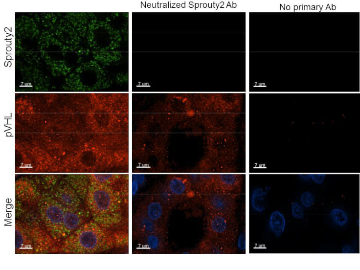
Anti-Sprouty-2 (RABBIT) Antibody
Sprouty-2 Antibody
- SPECIFICATION
- CITATIONS
- PROTOCOLS
- BACKGROUND

| Host | Rabbit |
|---|---|
| Conjugate | Unconjugated |
| Target Species | Human |
| Reactivity | Human |
| Clonality | Polyclonal |
Application
| WB, IHC, E, I, LCI |
| Application Note | Anti-Sprouty-2 antibody has been tested by ELISA, Immunofluorescence, Immunohistochemistry, and is suitable for western blotting. Specific conditions for reactivity should be optimized by the end user. |
| Physical State | Liquid (sterile filtered) |
| Buffer | 0.02 M Potassium Phosphate, 0.15 M Sodium Chloride, pH 7.2 |
| Immunogen | Anti-Sprouty-2 affinity purified antibody was purified from monospecific rabbit antiserum prepared via repeated immunizations with peptide corresponding to an internal region of human sprouty-2. |
| Preservative | 0.01% (w/v) Sodium Azide |
| Gene ID | 10253 |
|---|---|
| Other Names | 10253 |
| Purity | Anti-Sprouty-2 was affinity purified from monospecific antiserum by immunoaffinity chromatography. Sprouty-2 antibody reacts with human Sprouty 2 protein. A BLAST analysis was used to suggest reactivity with Sprouty 2 from human, monkey, and orangutan based on a 100% homology with the immunizing sequence. Cross-reactivity with Sprouty2 from other sources has not been determined. |
| Storage Condition | Store vial at -20° C prior to opening. Aliquot contents and freeze at -20° C or below for extended storage. Avoid cycles of freezing and thawing. Centrifuge product if not completely clear after standing at room temperature. This product is stable for several weeks at 4° C as an undiluted liquid. Dilute only prior to immediate use. |
| Precautions Note | This product is for research use only and is not intended for therapeutic or diagnostic applications. |
| Name | SPRY2 |
|---|---|
| Function | Antagonist of fibroblast growth factor (FGF) pathways via inhibition of FGF-mediated phosphorylation of ERK1/2 (By similarity). Thereby acts as an antagonist of FGF-induced retinal lens fiber differentiation, may inhibit limb bud outgrowth and may negatively modulate respiratory organogenesis (By similarity). Inhibits TGFB- induced epithelial-to-mesenchymal transition in retinal lens epithelial cells (By similarity). Inhibits CBL/C-CBL-mediated EGFR ubiquitination (PubMed:17974561). |
| Cellular Location | Cytoplasm, cytoskeleton. Cell projection, ruffle membrane. Note=Associated with microtubules in unstimulated cells but is translocated to the membrane ruffles in cells stimulated with EGF (epidermal growth factor) |

Thousands of laboratories across the world have published research that depended on the performance of antibodies from Abcepta to advance their research. Check out links to articles that cite our products in major peer-reviewed journals, organized by research category.
info@abcepta.com, and receive a free "I Love Antibodies" mug.

Provided below are standard protocols that you may find useful for product applications.
Background
Sprouty was originally identified as an inhibitor of Drosophila epidermal growth factor (EGF) and fibroblast growth factor (FGF) receptor signaling during tracheal development. Four isoforms of mammalian sprouty are known (Spry1-Spry4). The role of the mammalian analogs has not been clearly elucidated although it is believed that human Sprouty-2 (hSpry2) may be an inhibitor of cellular migration and proliferation. Spry2 and Spry4 show considerable sequence homology between human and mouse at the C-terminus of the protein. Significant sequence divergence occurs at the N-terminus. Spry2 appears to be abundant in brain, lung and heart tissue, with lesser amounts found in kidney and skeletal muscle.
If you have used an Abcepta product and would like to share how it has performed, please click on the "Submit Review" button and provide the requested information. Our staff will examine and post your review and contact you if needed.
If you have any additional inquiries please email technical services at tech@abcepta.com.
Ordering Information
Other Products
Shipping Information
