


 Foundational characteristics of cancer include proliferation, angiogenesis, migration, evasion of apoptosis, and cellular immortality. Find key markers for these cellular processes and antibodies to detect them.
 The SUMOplot™ Analysis Program predicts and scores sumoylation sites in your protein. SUMOylation is a post-translational modification involved in various cellular processes, such as nuclear-cytosolic transport, transcriptional regulation, apoptosis, protein stability, response to stress, and progression through the cell cycle.
 The Autophagy Receptor Motif Plotter predicts and scores autophagy receptor binding sites in your protein. Identifying proteins connected to this pathway is critical to understanding the role of autophagy in physiological as well as pathological processes such as development, differentiation, neurodegenerative diseases, stress, infection, and cancer.
HGS Antibody (monoclonal) (M01)
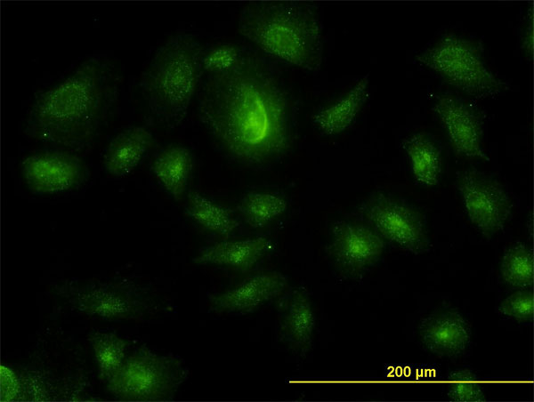
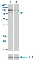
Mouse monoclonal antibody raised against a partial recombinant HGS.
- SPECIFICATION
- CITATIONS
- PROTOCOLS
- BACKGROUND

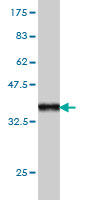
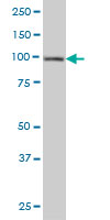
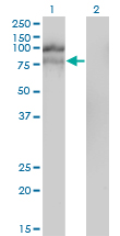
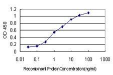
Application
| WB, IF, E |
|---|---|
| Primary Accession | O14964 |
| Other Accession | BC003565 |
| Reactivity | Human |
| Host | mouse |
| Clonality | Monoclonal |
| Isotype | IgG1 Kappa |
| Clone Names | 6D11 |
| Calculated MW | 86192 Da |
| Gene ID | 9146 |
|---|---|
| Other Names | Hepatocyte growth factor-regulated tyrosine kinase substrate, Hrs, Protein pp110, HGS, HRS |
| Target/Specificity | HGS (AAH03565, 513 a.a. ~ 612 a.a) partial recombinant protein with GST tag. MW of the GST tag alone is 26 KDa. |
| Dilution | WB~~1:500~1000 IF~~1:50~200 E~~N/A |
| Format | Clear, colorless solution in phosphate buffered saline, pH 7.2 . |
| Storage | Store at -20°C or lower. Aliquot to avoid repeated freezing and thawing. |
| Precautions | HGS Antibody (monoclonal) (M01) is for research use only and not for use in diagnostic or therapeutic procedures. |

Thousands of laboratories across the world have published research that depended on the performance of antibodies from Abcepta to advance their research. Check out links to articles that cite our products in major peer-reviewed journals, organized by research category.
info@abcepta.com, and receive a free "I Love Antibodies" mug.

Provided below are standard protocols that you may find useful for product applications.
References
1.The Class III Kinase Vps34 Promotes T Lymphocyte Survival through Regulating IL-7R? Surface Expression.McLeod IX, Zhou X, Li QJ, Wang F, He YW.J Immunol. 2011 Nov 15;187(10):5051-61. Epub 2011 Oct 21.
If you have used an Abcepta product and would like to share how it has performed, please click on the "Submit Review" button and provide the requested information. Our staff will examine and post your review and contact you if needed.
If you have any additional inquiries please email technical services at tech@abcepta.com.
Ordering Information
Other Products
Shipping Information
