


 Foundational characteristics of cancer include proliferation, angiogenesis, migration, evasion of apoptosis, and cellular immortality. Find key markers for these cellular processes and antibodies to detect them.
 The SUMOplot™ Analysis Program predicts and scores sumoylation sites in your protein. SUMOylation is a post-translational modification involved in various cellular processes, such as nuclear-cytosolic transport, transcriptional regulation, apoptosis, protein stability, response to stress, and progression through the cell cycle.
 The Autophagy Receptor Motif Plotter predicts and scores autophagy receptor binding sites in your protein. Identifying proteins connected to this pathway is critical to understanding the role of autophagy in physiological as well as pathological processes such as development, differentiation, neurodegenerative diseases, stress, infection, and cancer.
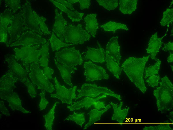
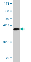
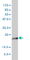
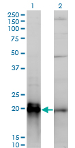
KRAS Antibody (monoclonal) (M01)
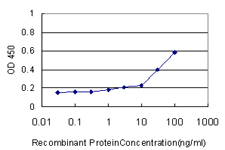
Mouse monoclonal antibody raised against a full length recombinant KRAS.
- SPECIFICATION
- CITATIONS: 1
- PROTOCOLS
- BACKGROUND

Application
| WB, IF, E |
|---|---|
| Primary Accession | P01116 |
| Other Accession | BC013572 |
| Reactivity | Human |
| Host | Mouse |
| Clonality | Monoclonal |
| Isotype | IgG1 Kappa |
| Clone Names | S2 |
| Calculated MW | 21656 Da |
| Gene ID | 3845 |
|---|---|
| Other Names | GTPase KRas, K-Ras 2, Ki-Ras, c-K-ras, c-Ki-ras, GTPase KRas, N-terminally processed, KRAS, KRAS2, RASK2 |
| Target/Specificity | KRAS (AAH13572, 1 a.a. ~ 188 a.a) full-length recombinant protein with GST tag. MW of the GST tag alone is 26 KDa. |
| Dilution | WB~~1:500~1000 IF~~1:50~200 E~~N/A |
| Format | Clear, colorless solution in phosphate buffered saline, pH 7.2 . |
| Storage | Store at -20°C or lower. Aliquot to avoid repeated freezing and thawing. |
| Precautions | KRAS Antibody (monoclonal) (M01) is for research use only and not for use in diagnostic or therapeutic procedures. |

Provided below are standard protocols that you may find useful for product applications.
Background
This gene, a Kirsten ras oncogene homolog from the mammalian ras gene family, encodes a protein that is a member of the small GTPase superfamily. A single amino acid substitution is responsible for an activating mutation. The transforming protein that results is implicated in various malignancies, including lung adenocarcinoma, mucinous adenoma, ductal carcinoma of the pancreas and colorectal carcinoma. Alternative splicing leads to variants encoding two isoforms that differ in the C-terminal region.
References
1.Evidence for aldosterone-dependent growth of renal cell carcinoma.King S, Bray S, Galbraith S, Christie L, Fleming SInt J Exp Pathol. 2014 May 7. doi: 10.1111/iep.12074.2.KRAS gene amplification in colorectal cancer and impact on response to EGFR-targeted therapy.Valtorta E, Misale S, Sartore-Bianchi A, Nagtegaal ID, Paraf F, Lauricella C, Dimartino V, Hobor S, Jacobs B, Ercolani C, Lamba S, Scala E, Veronese S, Laurent-Puig P, Siena S, Tejpar S, Mottolese M, Punt CJ, Gambacorta M, Bardelli A, Di Nicolantonio FInt J Cancer. 2013 Feb 12. doi: 10.1002/ijc.28106.3.Analysis of k-ras nuclear expression in fibroblasts and mesangial cells.Fuentes-Calvo I, Blazquez-Medela AM, Santos E, Lopez-Novoa JM, Martinez-Salgado C.PLoS One. 2010 Jan 14;5(1):e8703.
If you have used an Abcepta product and would like to share how it has performed, please click on the "Submit Review" button and provide the requested information. Our staff will examine and post your review and contact you if needed.
If you have any additional inquiries please email technical services at tech@abcepta.com.
Ordering Information
Other Products
Shipping Information
